
Page 254 - Feline diagnostic imaging
P. 254
258 14 Feline Pulmonary Disease
(a) (b)
(c)
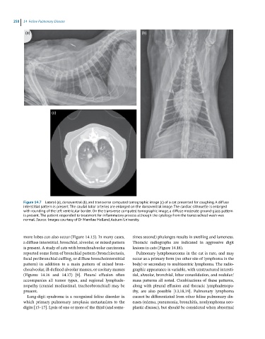
Figure 14.7 Lateral (a), dorsoventral (b), and transverse computed tomographic image (c) of a cat presented for coughing. A diffuse
interstitial pattern is present. The caudal lobar arteries are enlarged on the dorsoventral image. The cardiac silhouette is enlarged
with rounding of the left ventricular border. On the transverse computed tomographic image, a diffuse moderate ground-glass pattern
is present. The patient responded to treatment for inflammatory process although the cytology from the transtracheal wash was
normal. Source: Images courtesy of Dr Merrilee Holland, Auburn University.
more lobes can also occur (Figure 14.13). In many cases, times second) phalanges results in swelling and lameness.
a diffuse interstitial, bronchial, alveolar, or mixed pattern Thoracic radiographs are indicated in aggressive digit
is present. A study of cats with bronchoalveolar carcinoma lesions in cats (Figure 14.18).
reported some form of bronchial pattern (bronchiectasis, Pulmonary lymphosarcoma in the cat is rare, and may
focal peribronchial cuffing, or diffuse bronchointerstitial occur as a primary form (no other site of lymphoma in the
pattern) in addition to a main pattern of mixed bron- body) or secondary to multicentric lymphoma. The radio-
choalveolar, ill‐defined alveolar masses, or cavitary masses graphic appearance is variable, with unstructured intersti-
(Figures 14.16 and 14.17) [9]. Pleural effusion often tial, alveolar, bronchial, lobar consolidation, and nodular/
accompanies all tumor types, and regional lymphade- mass patterns all noted. Combinations of these patterns,
nopathy (cranial mediastinal, tracheobronchial) may be along with pleural effusion and thoracic lymphadenopa-
present. thy, are also possible [13,18,19]. Pulmonary lymphoma
Lung‐digit syndrome is a recognized feline disorder in cannot be differentiated from other feline pulmonary dis-
which primary pulmonary neoplasia metastasizes to the eases (edema, pneumonia, bronchitis, nonlymphoma neo-
digits [15–17]. Lysis of one or more of the third (and some- plastic disease), but should be considered when abnormal