
Page 259 - Feline diagnostic imaging
P. 259
14.4 Bronchial Disease 263
(a) (b)
(c)
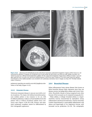
Figure 14.16 Lateral (a) and ventrodorsal (b) and postcontrast transverse computed tomographic image (c) of the thorax of a cat
presented for episodes of dyspnea. An ill-defined mass is seen within the left caudal lung lobe and right middle lung lobe. The
remaining lung has a diffuse bronchointerstitial pattern. On the computed tomographic image, there is diffuse increase in ground-
glass opacities with variably sized soft tissue nodules/masses. A soft tissue mass visualized in the left caudal lung lobe appears
bronchocentric. Fine needle aspiration was consistent with pulmonary carcinoma. Source: Images courtesy of Dr Merrilee Holland,
Auburn University.
pulmonary opacities are noted in cats with lymphoma else- 14.4 Bronchial Disease
where in the body (Figure 14.19).
Feline inflammatory lower airway disease (also known as
feline bronchial disease) likely represents more than one
14.3.2 Metastatic Disease
disease process, including feline asthma and chronic bron-
Pulmonary metastatic disease in cats can occur with a vari- chitis. Bronchiolar disease is being recognized more often
ety of patterns. The classic well‐defined interstitial pulmo- because of the increased use of computed tomography [21].
nary nodular pattern is less common in the cat, with Feline asthma, or allergic bronchial asthma, is a hyper-
ill‐defined pulmonary nodules or focal/multifocal alveolar sensitivity reaction characterized by eosinophilic airway
pattern with or without ill‐defined nodules more com- inflammation and bronchoconstriction. Chronic bronchitis
monly seen (Figure 14.20) [8,13,20]. Primary and meta- is better characterized by a neutrophilic inflammation with
static pulmonary neoplasia cannot be differentiated by edema and hypertrophy of the respiratory mucosa, with
their radiographic appearance. increased mucus production [22–25]. The radiographic