
Page 243 - Feline diagnostic imaging
P. 243
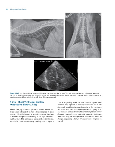
246 13 Acquired Heart Disease
(b)
(a)
(c)
Figure 13.43 A 15-year-old cat presented following chemotherapy due to fever. Thoracic lateral (a) and ventrodorsal (b) images of
the thorax show mild rounding and elongation of the left ventricular border. On the 2D image (c), the septal leaflet of the mitral valve
was thickened and hyperechoic (arrow). Endocarditis was suspected.
13.19 Right Ventricular Outflow 1.7 m/s originating from the infundibular region. This
Obstruction (Figure 13.46) murmur was reported to decrease when the heart rate
decreased, as did the increased velocity in the right ven
Before 1990, up to 20% of systolic murmurs had no ana tricular outflow tract. The majority of cats in a group of 51
tomic cause identified on the echocardiogram. A more had no other cardiac abnormality and the right ventricular
recently identified cause of systolic murmur has been chamber appeared normal on the 2D image. In 10/51 cats,
attributed to a dynamic narrowing of the right ventricular the echocardiogram was repeated in one year and found no
outflow tract. This appears as turbulent flow in the right change, suggesting a benign process without progression
ventricular outflow tract during systole greater or equal to [38,39].