
Page 235 - Feline diagnostic imaging
P. 235
238 13 Acquired Heart Disease
(a) (b)
(c)
(d) (e)
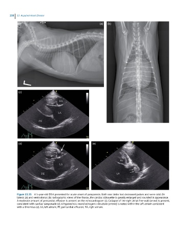
Figure 13.35 A 5-year-old DSH presented for acute onset of paraparesis. Both rear limbs had decreased pulses and were cold. On
lateral (a) and ventrodorsal (b) radiographic views of the thorax, the cardiac silhouette is greatly enlarged and rounded in appearance.
A moderate amount of pericardial effusion is present on the echocardiogram (c). Collapse of the right atrial free wall (arrow) is present,
consistent with cardiac tamponade (d). A hyperechoic round echogenic structure (arrows) is noted within the left atrium consistent
with a thrombus (e). LA, left atrium; PE, pericardial effusion; RA, right atrium.