
Page 230 - Feline diagnostic imaging
P. 230
13.14 Arteriotcromboembolism 233
(a)
(b)
(c)
(d) (e)
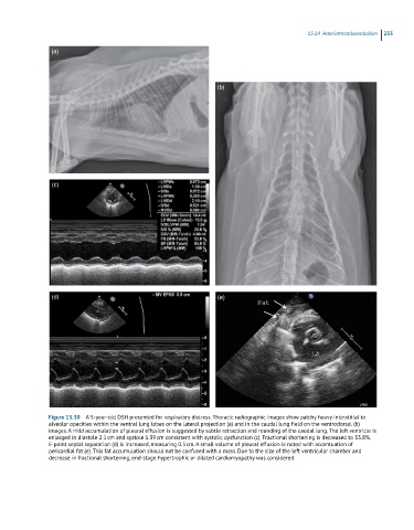
Figure 13.30 A 5-year-old DSH presented for respiratory distress. Thoracic radiographic images show patchy heavy interstitial to
alveolar opacities within the ventral lung lobes on the lateral projection (a) and in the caudal lung field on the ventrodorsal (b)
images. A mild accumulation of pleural effusion is suggested by subtle retraction and rounding of the caudal lung. The left ventricle is
enlarged in diastole 2.1 cm and systole 1.39 cm consistent with systolic dysfunction (c). Fractional shortening is decreased to 33.8%.
E-point septal separation (d) is increased, measuring 0.5 cm. A small volume of pleural effusion is noted with accentuation of
pericardial fat (e). This fat accumulation should not be confused with a mass. Due to the size of the left ventricular chamber and
decrease in fractional shortening, end-stage hypertrophic or dilated cardiomyopathy was considered.