
Page 223 - Feline diagnostic imaging
P. 223
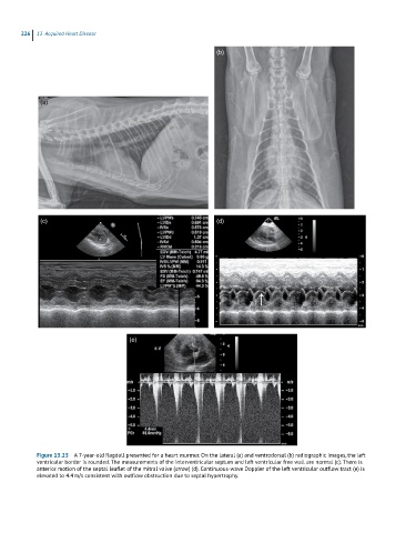
226  13  Acquired Heart Disease

                                                                 (b)


             (a)


             (c)                                                 (d)


                                       (e)


            Figure 13.23  A 7-year-old Ragdoll presented for a heart murmur. On the lateral (a) and ventrodorsal (b) radiographic images, the left
            ventricular border is rounded. The measurements of the interventricular septum and left ventricular free wall are normal (c). There is
            anterior motion of the septal leaflet of the mitral valve (arrow) (d). Continuous-wave Doppler of the left ventricular outflow tract (e) is
            elevated to 4.4 m/s consistent with outflow obstruction due to septal hypertrophy.