
Page 217 - Feline diagnostic imaging
P. 217
220 13 Acquired Heart Disease
(a) (c)
(b) (d)
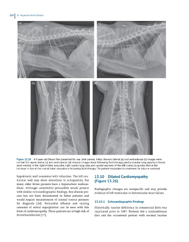
Figure 13.18 A 9-year-old Devon Rex presented for rear limb paresis. Initial thoracic lateral (a) and ventrodorsal (b) images were
normal. On repeat lateral (c) and ventrodorsal (d) thoracic images made following fluid therapy, patchy alveolar lung opacity is found,
most notably in the right middle lung lobe, right caudal lung lobe, and caudal segment of the left cranial lung lobe. Notice the
increase in size of the cranial lobar vasculature following fluid therapy. The patient responded to treatment for volume overload.
hypokinetic wall consistent with infarction. The left ven 13.10 Dilated Cardiomyopathy
tricular wall may show alterations in echogenicity, but (Figure 13.26)
many older feline patients have a hyperechoic endocar
dium. Although constrictive pericarditis would present Radiographic changes are nonspecific and may provide
with similar echocardiographic findings, this disease pro evidence of left ventricular or biventricular heart failure.
cess has not been documented in feline patients and
would require measurement of central venous pressure
for diagnosis [24]. Pericardial effusion and varying 13.10.1 Echocardiographic Findings
amounts of mitral regurgitation can be seen with this Historically, taurine deficiency in commercial diets was
form of cardiomyopathy. These patients are at high risk of implicated prior to 1987. Patients fed a nontraditional
thromboembolism [17]. diet and the occasional patient with normal taurine