
Page 212 - Feline diagnostic imaging
P. 212
13.7 Hypertropcic Cardiomyopatcy 215
(b)
(a)
(c) (d)
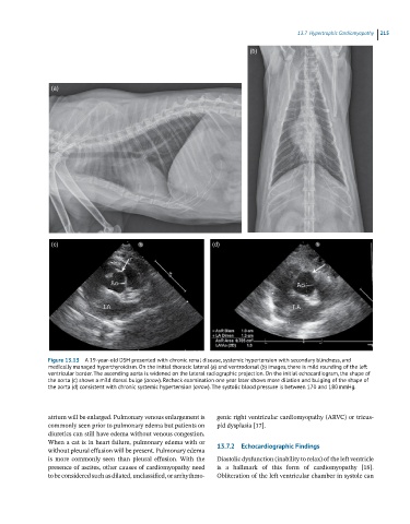
Figure 13.13 A 19-year-old DSH presented with chronic renal disease, systemic hypertension with secondary blindness, and
medically managed hyperthyroidism. On the initial thoracic lateral (a) and ventrodorsal (b) images, there is mild rounding of the left
ventricular border. The ascending aorta is widened on the lateral radiographic projection. On the initial echocardiogram, the shape of
the aorta (c) shows a mild dorsal bulge (arrow). Recheck examination one year later shows more dilation and bulging of the shape of
the aorta (d) consistent with chronic systemic hypertension (arrow). The systolic blood pressure is between 170 and 180 mmHg.
atrium will be enlarged. Pulmonary venous enlargement is genic right ventricular cardiomyopathy (ARVC) or tricus
commonly seen prior to pulmonary edema but patients on pid dysplasia [17].
diuretics can still have edema without venous congestion.
When a cat is in heart failure, pulmonary edema with or 13.7.2 Echocardiographic Findings
without pleural effusion will be present. Pulmonary edema
is more commonly seen than pleural effusion. With the Diastolic dysfunction (inability to relax) of the left ventricle
presence of ascites, other causes of cardiomyopathy need is a hallmark of this form of cardiomyopathy [18].
to be considered such as dilated, unclassified, or arrhythmo Obliteration of the left ventricular chamber in systole can