
Page 208 - Feline diagnostic imaging
P. 208
13.4 Hypertcyroidism 211
hypertrophic cardiomyopathy (HCM). Increased concen guished from cats in heart failure by surveyed practitioners
trations of cTnI are not specific for cardiomyopathy and with a 90% accuracy [7]. Neither cardiac troponin I nor NT
can be seen with mitral dysplasia, trauma, arrhythmia, pro‐B natriuretic peptide could distinguish cardiac changes
myocarditis, and congestive heart failure [4]. Different lev from HCM [8].
els of serum atrial and brain natriuretic peptides (BNP) can
be seen with HCM with and without failure in normal cats.
These findings support the use of this biomarker for screen 13.4 Hyperthyroidism
ing of cats for cardiac disease [5].
In a more recent study, a more refined test using plasma Hyperthyroidism is the most common geriatric endo
N‐terminal fragments of BNP could distinguish between crinopathy in cats. Cats with hyperthyroidism have been
normal cats and cats with occult heart disease [6]. Using found to have concurrent systemic hypertension in 5–22%
N‐terminal pro‐B natriuretic peptide along with standard of cases [9]. The typical thoracic radiographic changes
diagnostic testing such as thoracic radiographs, cats in res associated with hyperthyroidism include left ventricular
piratory distress due to noncardiac causes could be distin and left atrial enlargement (Figure 13.10a,b). In untreated
(a) (b)
(c)
(d)
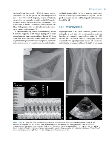
Figure 13.10 A 12-year-old DSH cat recently diagnosed with hyperthyroidism presented for treatment. Both lateral (a) and
ventrodorsal (b) projections of the thorax show enlargement of the left ventricle. The pulmonary parenchyma is normal. An
echocardiogram made on presentation shows thickening of the interventricular septum at 0.7 cm and left ventricular free wall at
0.6 cm in diastole. The left ventricular chamber is decreased in systole with an elevation of fractional shortening to 73% (c). The left
atrium is enlarged, measuring 1.76 cm from the right parasternal short axis view (d).