
Page 203 - Feline diagnostic imaging
P. 203
206 13 Acquired Heart Disease
(b)
(a)
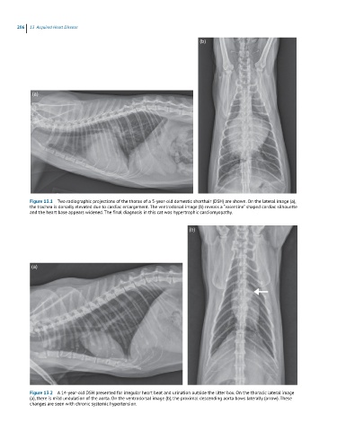
Figure 13.1 Two radiographic projections of the thorax of a 5-year-old domestic shorthair (DSH) are shown. On the lateral image (a),
the trachea is dorsally elevated due to cardiac enlargement. The ventrodorsal image (b) reveals a “valentine” shaped cardiac silhouette
and the heart base appears widened. The final diagnosis in this cat was hypertrophic cardiomyopathy.
(b)
(a)
Figure 13.2 A 14-year-old DSH presented for irregular heart beat and urination outside the litter box. On the thoracic lateral image
(a), there is mild undulation of the aorta. On the ventrodorsal image (b), the proximal descending aorta bows laterally (arrow). These
changes are seen with chronic systemic hypertension.