
Page 198 - Feline diagnostic imaging
P. 198
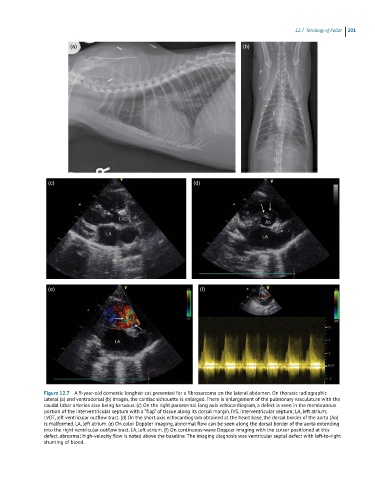
12.7 Tetralogy lof raale 201
(a) (b)
(c) (d)
(e) (f)
Figure 12.7 A 9-year-old domestic longhair cat presented for a fibrosarcoma on the lateral abdomen. On thoracic radiographic
lateral (a) and ventrodorsal (b) images, the cardiac silhouette is enlarged. There is enlargement of the pulmonary vasculature with the
caudal lobar arteries also being tortuous. (c) On the right parasternal long axis echocardiogram, a defect is seen in the membranous
portion of the interventricular septum with a “flap” of tissue along its dorsal margin. IVS, interventricular septum; LA, left atrium;
LVOT, left ventricular outflow tract. (d) On the short axis echocardiogram obtained at the heart base, the dorsal border of the aorta (Ao)
is malformed. LA, left atrium. (e) On color Doppler imaging, abnormal flow can be seen along the dorsal border of the aorta extending
into the right ventricular outflow tract. LA, left atrium. (f) On continuous-wave Doppler imaging with the cursor positioned at this
defect, abnormal high-velocity flow is noted above the baseline. The imaging diagnosis was ventricular septal defect with left-to-right
shunting of blood.