
Page 192 - Feline diagnostic imaging
P. 192
12.5 atent Ductus Arteriosus 195
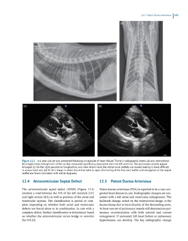
(b)
(a)
(c) (d)
Figure 12.2 A 6-year-old cat was presented following an episode of heart failure. Thoracic radiographic lateral (a) and ventrodorsal
(b) images show enlargement of the cardiac silhouette specifically associated with the left ventricle. The pulmonary vessels appear
enlarged. (c) On the right parasternal longitudinal axis view shown here, the mitral valve leaflets are closed, making it more difficult
to assess their size. (d) On this image in which the mitral valve is open, shortening of the free wall leaflet and elongation of the septal
leaflet are found consistent with mitral dysplasia.
12.4 Atrioventricular Septal Defect 12.5 Patent Ductus Arteriosus
The atrioventricular septal defect (AVSD) (Figure 12.9) Patent ductus arteriosus (PDA) is reported to be a rare con-
involves a void between the IVS of the left ventricle (LV) genital heart disease in cats. Radiographic changes are con-
and right atrium (RA) as well as portions of the atrial and sistent with a left atrial and ventricular enlargement. The
ventricular septum. The classification is partial or com- hallmark change, noted on the ventrodorsal image, is the
plete depending on whether both atrial and ventricular ductus bump due to focal dilation of the descending aorta.
defects are found alone or in combination. In cats with a At least one set of pulmonary vessels will demonstrate pul-
complete defect, further classification is determined based monary overcirculation with both arterial and venous
on whether the atrioventricular valves bridge or override enlargement. If untreated, left heart failure or pulmonary
the IVS [3]. hypertension can develop. The key radiographic change