
Page 173 - Feline diagnostic imaging
P. 173
174 10 Normal Cardiovascular Imaging
the PV. As the PVs open and close, the spectral Doppler valve closes and before it opens again and be depicted below
image will be a waveform with laminar flow depicted the baseline on the spectral Doppler imaging.
below the baseline (Figure 10.37). A sharp line can be seen
when the valve opens and closes; however, this does not 10.7.6 Tricuspid Valve
depict insufficiency. The normal laminar flow at the PV in
cats is typically less than 1.2 m/s [12]. Insufficiency would Spectral Doppler of the TV appears similar to the MV with
be above the baseline after the valve closes and before it a decreased amplitude in the normal flow (Figure 10.40).
opens again. Insufficiency at this valve greater than 1.9 m/s
is consistent with pulmonary hypertension. If the color 10.7.7 Aortic Valve
Doppler is insensitive on your machine, then mapping of The aortic valve can be the most technically difficult valve to
the valve by using pulsed‐wave or continuous‐wave align properly for Doppler evaluation. With the footprint of
Doppler imaging in the right ventricular outflow tract will the probe in the same location, the angle of the probe may
isolate any insufficiency. Mapping means moving the box need to be steepened by pulling the handle toward the table
or cursor above the valve in the outflow tract, keeping in and caudally toward the body wall. The normal velocity at
mind theat insufficiency does not always go directly above the aortic valve is typically less than 1.2 m/s [12]. The velocity
the valves.
at the level of the aortic valve (Figure 10.41) and within the
LVOT (Figure 10.42) should be documented. If narrowing is
present in the LVOT due to hypertrophy of the IVS, continu-
10.7.5 Mitral Valve
ous‐wave Doppler should be used because higher velocities
On the left parasternal long axis view of the heart, the MV cannot be recorded with pulsed‐wave Doppler. Increased
and aortic valve are normally seen adjacent to each other, velocity in the LVOT is consistent with outflow obstruction
assisting in identification of left versus right heart. From the and can occur in older cats with asymmetric hypertrophic
observation deck, the blood flow moves from the LA cardiomyopathy or in a young cat with mitral dysplasia.
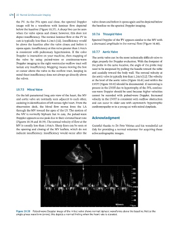
through the MV toward the apex of the LV. The motion of
the MV is normally biphasic but in cats, the pulsed‐wave
Doppler appears as one peak due to their elevated heart rate Acknowledgment
(Figures 10.38 and 10.39). The normal velocity of flow at the
MV is usually less than 1.0 m/s. Sharp lines can be seen at Grateful thanks to Dr Pete Velotas and his wonderful cat
the opening and closing of the MV leaflets, which do not Ody for providing a normal volunteer for acquiring these
indicate insufficiency. Insufficiency would occur after the echocardiographic images.
Figure 10.38 Pulsed-wave Doppler image of the mitral valve shows normal biphasic waveforms above the baseline. Notice the
single-phase waveform (arroe); this depicts a normal finding when the heart rate is elevated.