
Page 569 - Veterinary Immunology, 10th Edition
P. 569
VetBooks.ir
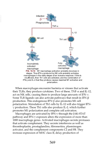
FIG. 18.14 M1 macrophage activation probably develops in
stages. Thus IFN-γ produced by NK cells probably activates
macrophages in the early stages of an immune response. If this is
insufficient, then Th1 cells are activated, and the combination of
IFN-γ and IL-2 that they produce causes maximal M1 activation and
polarization.
When macrophages encounter bacteria or viruses that activate
their TLRs, they produce cytokines. Two of these, TNF-α and IL-12,
act on NK cells, causing them to produce large amounts of IFN-γ.
Some TLR ligands can also activate pathways that result in IFN-β
production. This endogenous IFN-β also promotes M1 cell
polarization. Stimulation of Th1 cells by IL-12 will also trigger IFN-
γ production. These Th1 cells also produce IL-2, which further
promotes M1 polarization and complete cell activation.
Macrophages are activated by IFN-γ through the JAK-STAT
pathway and IFN-γ exposure alters the expression of more than
1000 macrophage genes. Activated macrophages secrete proteases
that activate complement. They secrete interferons as well as
thromboplastin, prostaglandins, fibronectins, plasminogen
activator, and the complement components C2 and FB. They
increase expression of MHC class II, delay production of
569