
Page 573 - Veterinary Immunology, 10th Edition
P. 573
tuberculosis. Thus mycobacteria that enter the lungs are readily
VetBooks.ir phagocytosed by alveolar macrophages that then mount a
respiratory burst and secrete proinflammatory cytokines. These
cytokines act on NK cells, triggering IFN-γ production and limited
macrophage activation. This rapid response can slow mycobacterial
growth significantly. Nevertheless, these macrophages cannot
destroy the bacteria by these mechanisms alone. After several days,
however, recruitment of T cells occurs. The T cells are stimulated by
mycobacteria-infected dendritic cells secreting IL-12, TNF-α, and
IFN-α. In response, the Th1 cells secrete more IFN-γ and fully
activate the macrophages (Table 18.2). In most individuals, this
activation to the M1 level is sufficient to control the infection.
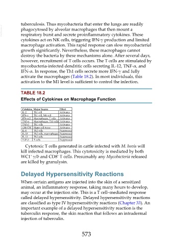
TABLE 18.2
Effects of Cytokines on Macrophage Function
Cytokine Major Source Effect
IL-2 Th1 cell Activates
IFN-γ Th1 cell, NK cell Activates
IFN-α/β Macrophages, T cells Activates
TNF-α Macrophages, Th1 cells Activates
TNF-β Th1 cells Activates
GM-CSF Many cell types Activates
IL-4 Th2 cells Suppresses
IL-10 Th2 cells, macrophages Suppresses
IL-13 Th2 cells Suppresses
TGF-β T cells Suppresses
Cytotoxic T cells generated in cattle infected with M. bovis will
kill infected macrophages. This cytotoxicity is mediated by both
+
+
WC1 γ/δ and CD8 T cells. Presumably any Mycobacteria released
are killed by granulysin.
Delayed Hypersensitivity Reactions
When certain antigens are injected into the skin of a sensitized
animal, an inflammatory response, taking many hours to develop,
may occur at the injection site. This is a T cell–mediated response
called delayed hypersensitivity. Delayed hypersensitivity reactions
are classified as type IV hypersensitivity reactions (Chapter 33). An
important example of a delayed hypersensitivity reaction is the
tuberculin response, the skin reaction that follows an intradermal
injection of tuberculin.
573