Page 570 - Veterinary Immunology, 10th Edition
P. 570
phagosomal proteases, and promote antigen loading onto MHC
  VetBooks.ir  molecules, so enhancing antigen presentation. M1 macrophages are

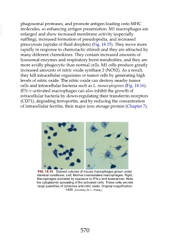
               enlarged and show increased membrane activity (especially
               ruffling), increased formation of pseudopodia, and increased

               pinocytosis (uptake of fluid droplets) (Fig. 18.15). They move more
               rapidly in response to chemotactic stimuli and they are attracted by
               many different chemokines. They contain increased amounts of
               lysosomal enzymes and respiratory burst metabolites, and they are

               more avidly phagocytic than normal cells. M1 cells produce greatly
               increased amounts of nitric oxide synthase 2 (NOS2). As a result,
               they kill intracellular organisms or tumor cells by generating high
               levels of nitric oxide. The nitric oxide can destroy nearby tumor

               cells and intracellular bacteria such as L. monocytogenes (Fig. 18.16).
               IFN-γ-activated macrophages can also inhibit the growth of
               intracellular bacteria by down-regulating their transferrin receptors
               (CD71), degrading ferroportin, and by reducing the concentration

               of intracellular ferritin, their major iron storage protein (Chapter 7).


                            FIG. 18.15  Stained cultures of mouse macrophages grown under
                           identical conditions: Left, Normal unstimulated macrophages. Right,
                           Macrophages activated by exposure to IFN-γ and acemannan. Note
                           the cytoplasmic spreading of the activated cells. These cells secrete
                           large quantities of cytokines and nitric oxide. Original magnification
                                               ×400. (Courtesy Dr. L. Zhang.)


                                                         570

