
Page 10 - GP Spring 2024
P. 10
Maxillary Sinusitis of Dental Origin (MSDO): A Case Report
Author: Joseph DiDonato, III, DDS, MBA, FAGD
Maxillary Sinusitis of Dental Origin sional radiographs is that the focal point symptoms, it wasn’t until the advent of the
(MSDO) or of Endodontic Origin (MSEO) may not be in the center of the intended tar- CBCT that we, as dentists, could see the di-
is a sinus infection that originates from a get, so a mild blurring occurs of objects in rect result- a thickening of the Schneiderian
necrotic tooth or a failed endodontically front of and behind the focal point. This can membrane. This is a mucositis that general-
treated tooth and subsequent infection in result in several diagnostic dilemmas. First, ly is not well depicted on periapical radio-
a posterior maxillary tooth. Some studies the full anatomy may not be captured and, graphs and only occasionally is demonstrat-
indicate that more than 40% of maxillary therefore, not seen. Second, there may be ed on a panoramic radiograph and is even
sinusitis cases are caused by a tooth infec- a complete miss of the pathology if the pa- more rarely diagnosed. Part of the problem
tion. The condition has been recognized in thology occurs outside the plane of focus. has been the ‘shared’ anatomy between den-
1
a position paper by the American Associa- Third, if the pathology results in a thinning tal and medical providers which has contrib-
tion of Endodontists: of naturally thin bone, as in the floor of the uted to the idea that each entity will diagnose
sinus, it may not demonstrate the disruption and treat disease as it is identified. The un-
“The relationship between dental infections of the cortical plate, and therefore, the pa- der-recognized and under-diagnosed status
and sinus disease is widely recognized in thology is not readily differentiated on the has contributed to cases that persist and go
both the dental and medical literature. De- two-dimensional image. untreated.
spite extensive scientific recognition and
reported high prevalence, periapical in- Within the last ten years, cone-beam com- Understanding the scale and scope of this
fection manifesting in the maxillary sinus puter tomography (CBCT) has demonstrated problem may require a more expansive
remains under-appreciated and frequently that the three-dimensional view is a superior review to include the otolaryngology lit-
goes undiagnosed by dentists, otolaryngol- modality for imaging odontogenic pathology erature to gain an appreciation of the di-
ogists, and radiologists alike, with its se- of the maxillary sinus. The CBCT provides agnostic criteria and treatment modalities
quelae often misdiagnosed as sinogenic si- focused imaging throughout the subject and that ENT specialists employ in these cases.
nusitis. Recognition of MSEO is critical as provides a unique view of the pathology However, the critical understanding is that
failure to identify and properly manage the caused by necrotic maxillary posterior teeth this is a disease caused by the dentition and
endodontic source pathology will result in to the sinus. Developing one plane of focus needs to be definitively resolved by either
the persistence of sinus disease, the failure on a maxillary molar is insufficient and mis- endodontic treatment or exodontia.
of medical sinus therapies, and the poten- leading for determining the condition of the
tial advancement to more serious or even tooth. One must image the entire root com- Presentation of a case:
life-threatening cranio-facial infections.” 2 plex for a complete survey. To reiterate, one A 65-year-old woman who had had regular
must be able to visualize all the apices of a dental care for over 30 years with extensive
Historical Background: maxillary tooth to describe the anatomy and full coverage restorations accomplished in
Maxillary sinusitis of dental origin (MSDO) radiographic condition of a tooth. the late 1990s presented to my office. She
was first recognized in 1943 by Bauer us- had no dental complaints and requested an
ing cadavers to find the direct extension of Patients with MSEO may experience typ- exam and oral hygiene visit. At the visit, a
dental disease into the sinus. Bauer dis- ical sino-nasal symptoms, including nasal standard survey of bitewings and panoramic
3
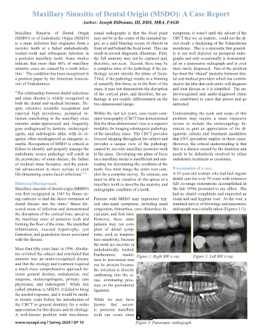
sected areas of infection and demonstrated congestion, rhinorrhea, retro rhinorrhea, fa- radiograph was initially taken (Figures 1-3).
the disruption of the cortical bone apical to cial pain, and foul odor.
the maxillary roots of posterior teeth and However, these same
forming the floor of the sinus. He identified patients may not com-
inflammation, mucosal hypertrophy, cyst plain of dental symp-
formation, and granulation tissue associated toms, such as tempera-
with the disease. ture sensitivity, because
the teeth are necrotic or
More than fifty years later, in 1996, Abraha- endodontically treated.
ms revisited the subject and concluded that Furthermore, tender- Figure 1. Right BW x-ray. Figure 2. Left BW x-ray.
sinusitis was an under-recognized disease ness to percussion may
and that the etiology and treatment required not be present because
a much more comprehensive approach be- the infection is directly
tween general dentists, endodontists, oral outflowing into the si-
surgeons, otolaryngologists, primary care nus, eliminating pres-
physicians, and radiologists. While this sure on the periodontal
4
called attention to MSDO, it failed to bring ligament.
the needed response, and it would be anoth-
er twenty years before the introduction of While we may have
the CBCT to general dentistry for a wider known that necrot-
appreciation for this disease and its etiology. ic posterior maxillary
A well-known problem with two-dimen- teeth can create sinus
www.nysagd.org l Spring 2024 l GP 10 Figure 3. Panoramic radiograph.