
Page 13 - GP Spring 2024
P. 13
A Technique for the Management of Deep and Subgingival Caries
Using a Bioclear Evolve Matrix
Authors: Daniel Abelson, DDS, and Arthur Volker, DDS, MSc, MSEd
Introduction rings can be selected based on tooth size and the need for grip
The composite class II restoration is a common yet technique-sen- strength, which is helpful when tissue is thick.
sitive procedure. Recreating interdental anatomy is one such chal-
lenge. Characteristics of ideal interdental anatomy include proxi- Case Presentation
mal surface with convex contour, contact with the adjacent tooth, A healthy 26-year-old male presented for routine re-care treatment
gingival margin seal, and correct height of the marginal ridge. and prophylaxis. A bitewing radiograph revealed the upper right
1
Failure to recreate ideal interdental anatomy may result in food second molar with a deep carious lesion extending below the
5
impaction (which can cause patient discomfort), gingivitis or peri- cemento-enamel junction. The patient was asymptomatic and the
odontal disease, marginal gap formation, or recurrent caries. tooth was vital. The tooth was initially restored with a standard
circumferential matrix, which resulted in an open contact. As food
Recurrent caries is the main cause of failure for both composite impaction became too bothersome for the patient, the decision was
and amalgam class II restorations, but the risk of recurrent caries made to re-do the restoration.
has been found to be higher in the composite than in amalgam.
2-4
Given the risk of recurrent caries, it is imperative to understand To improve results, several changes were implemented. First, a
the techniques necessary for long-lasting composite restorations. wooden wedge was used at the start of the procedure to create in-
In this regard, clinicians should be aware of clinical conditions terdental separation. Second, a gingivectomy was performed on
that present added levels of difficulty and should have knowledge the lingual papilla, which aided in both wedge placement and in
about which materials will help to address those difficulties. the ring’s ability to grip the wedge. Third, enameloplasty was per-
formed on the distal of the upper first molar to prevent the matrix
This case shows how various clinical conditions, including root from folding on itself. Fourth, the Bioclear sectional matrix system
proximity, subgingival caries, tooth anatomy such as emergence was used, which offered increased customization, including: an
profile and width, and thick palatal tissue, all converge to create “extra-long” wedge; “pink matrix,” that is broad bucco-lingually
restorative challenges. Given these clinical challenges, the case and has minimal proximal curvature; and a white twin-ring that has
also highlights the need for materials that offer improved custom- improved griping capability when the tissue is thick.
ization to recreate anatomy. In this regard, the Bioclear Evolve
Matrix System offers matrices that vary in three key dimensions: Figure 2. Pre-operative radiograph
emergence profile, width, and height (Figure 1). demonstrating an upper right second
molar with deep caries that extends
below the CEJ. Regarding the first
and second upper molars, the radio-
graph also shows two challenging
clinical conditions: root proximity
and interdental anatomy in which the
first molar’s distal surface creates a
slight concavity on the second molar’s
mesial surface.
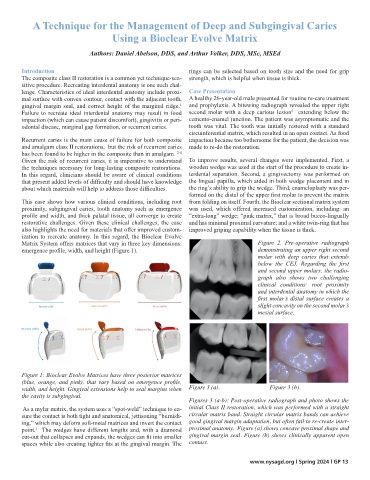
Figure 1: Bioclear Evolve Matrices have three posterior matrices
(blue, orange, and pink), that vary based on emergence profile,
width, and height. Gingival extensions help to seal margins when Figure 3 (a). Figure 3 (b).
the cavity is subgingival.
Figures 3 (a-b): Post-operative radiograph and photo shows the
As a mylar matrix, the system uses a “spot-weld” technique to en- initial Class II restoration, which was performed with a straight
sure the contact is both tight and anatomical, jettisoning “burnish- circular matrix band. Straight circular matrix bands can achieve
ing,” which may deform soft-metal matrices and invert the contact good gingival margin adaptation, but often fail to re-create inter-
point. The wedges have different lengths and, with a diamond proximal anatomy. Figure (a) shows concave proximal shape and
1
cut-out that collapses and expands, the wedges can fit into smaller gingival margin seal. Figure (b) shows clinically apparent open
spaces while also creating tighter fits at the gingival margin. The contact.
www.nysagd.org l Spring 2024 l GP 13