Page 11 - GP Spring 2024
P. 11

After the radiography was evaluated, the patient was asked about sinus problems.  The patient reported consistent, chronic sinus problems
      that were alternately treated by decongestants, antihistamines, and rarely antibiotics.  After much reflection, she remembered one occasion
      when she presented to her primary care physician with soreness in her cheek and was put on a course of antibiotics, which resolved the
      problem.  She was told it may be related to dry winter conditions.
                                                                               On the panoramic view, fine vertical opacifi-
                                                                               cations (halos) superior to the posterior max-
                                                                               illary  teeth,  bilaterally,  indicated  a  possible
                                                                               anomaly  and  additional  maxillary  posterior
                                                                               periapical  radiographs  were  taken  (Figures
                                                                               4, 5).  The radiographs revealed several teeth
                                                                               with full coverage crowns and extensive core
                                                                               build-ups.  The margins and cement seals were
                                                                               intact; there were no secondary caries; the pa-
                                                                               tient  reported  no  sensitivity  to  percussion  or
                                                                               chewing, no discomfort to hot or cold, and no
      Figure 4. Periapical x-ray, right side.  Figure 5. Periapical x-ray, left side.  sensation to electrical stimulation for vitality
                                                                               testing.  She also reported normal for her si-
                                                                               nus conditions, which meant typical seasonal
                                                                               allergies and post-nasal drip.  However, on the
                                                                               panoramic radiographs, the apical region of the
                                                                               maxillary  teeth  revealed  mild,  lace-like  halo
                                                                               arcs  with  discontinuous  boundaries  2-3  mm
                                                                               from the apices of teeth 2, 3, and 14, with the
                                                                               root apices as the center of the arc.
                                                                               The patient was advised that while the periapi-
                                                                               cal  and  panorex  images  did  not  demonstrate
                                                                               clear pathology, there was a high degree of sus-
                                                                               picion about the vitality of teeth #2, 3, and 14
                                                                               and their contribution to her sinus condition.  A
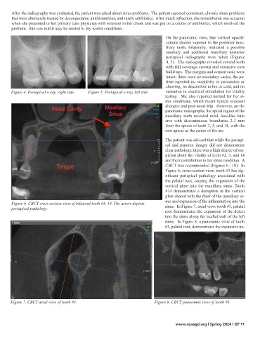
                                                                               CBCT was recommended (Figures 6 - 10).  In
                                                                               Figure 6, cross-section view, tooth #3 has sig-
                                                                               nificant  periapical  pathology  associated  with
                                                                               the palatal root, causing the expansion of the
                                                                               cortical  plate  into  the  maxillary  sinus. Tooth
                                                                               #14 demonstrates a disruption in the cortical
                                                                               plate shared with the floor of the maxillary si-
      Figure 6. CBCT cross section view of bilateral teeth #3, 14. The arrow depicts    nus and expansion of the inflammation into the
      periapical pathology.                                                    sinus.  In Figure 7, axial view, tooth #3, palatal
                                                                               root demonstrates the expansion of the defect
                                                                               into the sinus along the medial wall of the left
                                                                               sinus.  In Figure 8, a panoramic view of tooth
                                                                               #3, palatal root, demonstrates the expansive na-


      Figure 7. CBCT axial view of tooth #3.                              Figure 8. CBCT panoramic view of tooth #3.


                                                                                    www.nysagd.org l Spring 2024 l GP 11
   6   7   8   9   10   11   12   13   14   15   16