
Page 62 - parasitology for medical and clinical laboratoryprofessionals
P. 62
42 CHAPTER 3
apparently infected by the trophozoite or active stage
of metabolism in the life cycle of D. fragilis. The organ-
ism obtains nutrition by phagocytosis, as it extends the
pseudopodia and envelopes its prey, which it then pro-
ceeds to break down into simple nutrient materials. The
cytoplasm most often contains numerous food vacuoles
with ingested debris that may include bacteria. The con- Source: Centers for Disease Control and Prevention (CDC)
clusion whereby D. fragilis was classified as a nonpatho-
gen was based on its insatiable appetite for the normal
bacteria found in the gut rather than the intestinal and
other tissues of its host. Waste materials are eliminated
from the cell through digestive vacuoles by exocytosis.
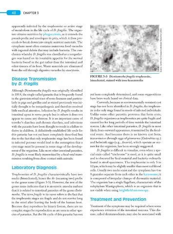
FIGURE 3-5 Dientamoeba fragilis trophozoite,
Disease Transmission binucleated, stained with iron-hematoxylin
by D. fragilis
Although Dientamoeba fragilis was originally identified
in 1918, the single-celled parasite that is frequently found yet been completely determined, and some suppositions
in the gastrointestinal tract of some humans and particu- have been made based on clinical data.
larly in pigs and gorillas and as stated previously was ini- Currently, because an environmentally resistant cyst
tially thought to be nonpathogenic and therefore received stage has not been identified in D. fragilis, the trophozo-
little medical attention. Infection by D. fragilis results in ite is the only stage found in stools of infected individuals.
intestinal upset in some people but in others it does not Unlike some other parasitic protozoa that form cysts,
appear to cause any distress. It is an important cause of D. fragilis organisms as trophozoites are quite fragile and
traveler’s diarrhea and chronic diarrhea, and infections cannot live for long periods of time outside the intestinal
with the parasite have been thought to result in a failure to system. Like other intestinal parasites, D. fragilis is most
thrive in children. A definitively established life cycle for likely, from outward appearance, transmitted by the fecal-
this parasite has not yet been completely described but oral route. And because there is no known cyst form,
due to the fact that only trophozoite stage has been found transmission through eggs of pinworms (Enterobius sp.)
in infected persons would lead to the assumption that a and helminth eggs (e.g., Ascaris), which operate as vec-
cyst stage must be present in some stage of the develop- tors for the organism, has been strongly suggested.
ment of the organism. Like most other intestinal parasites, D. fragilis is difficult to visualize, even when a spe-
D. fragilis is most likely transmitted by a fecal-oral trans- cial stain called “trichrome” is used, as it is quite small
mission resulting from close contact with animals. and is obscured by fecal material and bacteria ordinarily
found in stool specimens. The trophozoite is only 5 to
Laboratory Diagnosis 12 μm, which may be slightly smaller than most red blood
cells. Usually two nuclei exist and the cytoplasm has 4 to
Trophozoites of D. fragilis characteristically have two 8 granules separate from each other in the karyosome; it
nuclei (binucleated), hence the di- (meaning two) prefix is composed of irregular clumps of chromatin material.
to the genus name (Figure 3-5). However, the rest of the The organism has a single flagellum, characteristic of the
genus name indicates that it is an enteric amoeba and not subphylum Mastigophora, which is an organism that is
that it is related to intestinal parasites of the genus Enta- not visible when using brightfield microscopy.
moeba. The term fragile in its name refers to the fact that
the trophozoite stages are fragile and do not survive long Treatment and Prevention
in the stool after leaving the body of the human host.
Because they reproduce by binary fission, there are no Treatment of the symptoms may be required where some
complex stages for reproduction as are seen in other spe- experience irritation of the intestinal mucosa. This dis-
cies of parasites. But the life cycle of this parasite has not ease, called dientamoebiasis, may also be associated with