
Page 10 - textbook5chapters
P. 10
SURGICAL ANATOMY IN PELVIC
131GYNAECOLOGIC ONCOLOGY
Hugo Gaspar MD
Octavio Arencibia Sanchez, MD PhD
Jordi Ponce, MD PhD
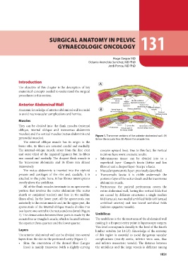
Introduction Transversalis
The objective of this chapter is the description of key fascia Medial Medial umbilical
anatomical concepts needed to understand the surgical Peritoneum umbilical ligament (uracaus)
procedures in this section. ligament
Transversus
Anterior Abdominal Wall abdominis musIcnlteernal oblique
Rectus abdominis
Anatomic knowledge of anterior abdominal wall is crucial Ventral layer of mucsle
to avoid neurovascular complications and hernias.
the rectus sheath External
Muscles
oblique muscle
They can be divided into: the flank muscles (external
oblique, internal oblique and transversus abdominis Transversalis
muscles) and the vertical muscles (rectus abdominis and
pyramidal muscles). fascia Linea alba Apneurosis of the
internal oblique muscle
The external oblique muscle has its origin in the Peritoneum Dorsal layer of
lower ribs, its fibers are oriented caudal and medially.
The internal oblique muscle arises from the iliac crest the rectus sheath
and outer third of the inguinal ligament but its fibers
run cranial and medially. The deepest flank muscle is Figure 1. Transverse sections of the anterior abdominal wall. (A)
the transversus abdominis and its fibers run almost Below the arcuate line. (B) Above the arcuate line.
transversely.
concave upward line). Due to this fact, the vertical
The rectus abdominis is inserted into the xiphoid incisions have worst cosmetic results.
process and cartilages of the ribs and, caudally, it is • Subcutaneous tissue: can be divided into to a
attached to the pubic bone. It has fibrous interruptions superficial layer- Camper’s fascia (fattier and less
mostly above the umbilicus. fibrous) and a deepest layer- Scarpa´s fascia.
• Muscularaponeurotic layer: previously described.
All of the flank muscles terminate in an aponeurotic • Transversalis fascia: it is visible underneath the
portion that involves the rectus abdominis (the rectus posterior layer of the rectus sheath and the transversus
sheath or conjoined tendon) and fuse in the midline abdominis muscle.
(linea alba). In the lower part, all the aponeurosis run • Peritoneum: the parietal peritoneum covers the
anteriorly to the rectus muscle and, in the upper part, the entire abdominal wall, having five vertical folds that
aponeurosis of the internal oblique muscle divides into are caused by different structures: a single median
two layers: one ventral to the rectus and one dorsal (Figure fold (uracus), two medial umbilical folds (obliterated
1). The demarcation between these parts is made by the umbilical arteries) and two lateral umbilical folds
arcuate line or Douglas’s arcade, which is located between (inferior epigastric vessels).
the superior three quarters and the lower quarter.
Umbilicus
Layers
The umbilicus is the thinnest area of the abdominal wall
The anterior abdominal wall can be divided into several making it a frequent entry point in laparoscopy surgery.
layers from the skin to the peritoneal cavity (Figure 1): This level corresponds dorsally to the level of the fourth
• Skin: the orientation of the dermal fiber (Langer lumbar vertebra (or L4-L5). Knowledge of the anatomy
of this region is essential to avoid dangerous vascular
lines) is mainly transverse (with a slightly curving complications (mainly aortic, inferior vena cava, iliac
and inferior mesenteric vessels). The distance between
the umbilicus and the large vessels is different among
1031