
Page 12 - textbook5chapters
P. 12
Surgical Anatomy in Pelvic Gynaecologic Oncology 1033
Figure 3. The female pelvis: the pelvic
bones, joints, ligaments and foramina.
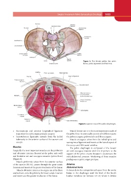
Figure 4. Superior view of the pelvic diaphragm.
• Sacrospinous and anterior longitudinal ligament: Muscle levator ani: it is the most important muscle of
important for pelvic organ prolapse surgery. the pelvic floor. Anatomically consists of 3 different parts:
the pubococcygeus, puborectalis and iliococcygeus.
• Sacrotuberous ligaments: extends from the ischial
tuberosity to the anterior surface of the sacrum and Muscle coccygeus: arises from the ischial spine and
coccyx. sacrospinous ligament and inserts at the lateral aspect of
the coccyx and fifth sacral vertebra.
Muscles
The pelvic diaphragm is composed of the levator
Surgically the most important muscles are the piriformis ani and coccygeus muscles and it is important to the
and obturator internus (located at the pelvic side wall) support of the pelvic viscera because it counteracts the
and levatador ani and coccygeus muscles (pelvic floor) intra-abdominal pressure. Weakening of these muscles
(Figure 4). predisposes to pelvic organ prolapse.
Muscle piriformis: arises from the anterior surface Arteries
of the sacrum (S2-S4), passes through the great sciatic Abdominal Aorta
foramen and inserts at the greater trochanter of the femur. It descends in the retroperitoneal space from the aortic
hiatus in the diaphragm until the level of the fourth
Muscle obturator internus: its origins are at the ilium lumbar vertebrae (or between L4-L5) where it divides
and ischium, exits the pelvis by the lesser sciatic foramen
and inserts and the greater trochanter of the femur.