
Page 14 - textbook5chapters
P. 14
Surgical Anatomy in Pelvic Gynaecologic Oncology 1035
Figure 8. Aortic bifurcation and left common iliac vein.
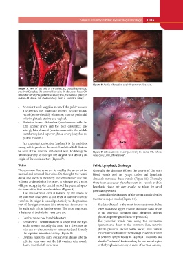
Figure 7. View of left side of the pelvis. BL, broad ligament; D,
pouch of Douglas; EIV, external iliac vein; OF obturator fossa; ON,
obturator nerve; PrS; pararectal space; PvS, Paravesical space; R,
rectum; U, uterus; UA, uterine artery; Umb. A, umbilical artery.
• Anterior trunk: supplies most of the pelvic viscera. Figure 9. Left renal vein crossing ventrally the aorta. IVC, inferior
The arteries are: umbilical; inferior vesical; middle vena cava; LRV, Left renal vein.
rectal (hemorrhoidal); obturator; internal pudendal;
inferior gluteal; uterine and vaginal. Pelvic Lymphatic Drainage
• Posterior trunk: iliolumbar (anastomoses with the Generally the drainage follows the course of the main
fifth lumbar artery and the deep circumflex iliac blood vessels and the lymph nodes and lymphatic
artery), lateral sacral (anastomoses with the middle channels surround these vessels (Figure 10). Normally,
sacral artery) and superior gluteal artery (supplies the there is an avascular plane between the vessels and the
gluteal muscles). lymphatic tissue but care should be taken for small
perforating vessels.
An important anatomical landmark is the umbilical
artery, which produces the medial umbilical fold that can Classically, the drainage of the cervix can de divided
be seen at the anterior abdominal wall. Following the into three major trunks (Figure 11):
umbilical artery to its origin the surgeon will identify the
origin of the uterine artery (Figure 7). - The lateral trunk is the most important route. It has
three branches (upper, middle and lower) and drains
Veins to the interiliac, common iliac, obturator, inferior
gluteal, superior gluteal and/or presacral;
The common iliac veins are formed by the union of the
internal and external iliac veins. On the right, the vein is - The posterior trunk runs along the uterosacral
dorsal and lateral to the artery. The left common iliac vein ligament and drain to the common iliac, superior
is dorsal and medial to the artery. It is longer and a more gluteal, presacral and/or aortic nodes. This route is
oblique, occupying the cranial part of the presacral space the anatomical basis for the findings in several studies
(in front of the first sacral vertebra) (Figure 8). of sentinel lymph nodes in “atypical” locations and
also the “rationale” for including the pre-sacral region
The inferior vena cava is formed by the union of in the lymphadenectomy in cases of cervical cancer;
the common iliac veins at the level of the fifth lumbar
vertebra. Its origin is located posteriorly to the proximal
part of the right common iliac artery and its courses on
the right side of the lumbar spine. The most important
tributaries of the inferior vena cava are:
• Lumbar veins: one for which artery.
• Renal veins: The left renal vein is longer than the right
and it crosses ventrally the aorta (in a few cases the
vein can be circumaortic or retroaortic) and dorsally
the superior mesenteric artery (Figure 9).
• Ovarian veins: the right ovarian vein drains into the
inferior vena cava but the left ovarian vein usually
drains into the left renal vein.