
Page 15 - textbook5chapters
P. 15
1036 Surgical Anatomy in Pelvic Gynaecologic Oncology
Figure 12. Nerves of the pelvic viscera.
Figure 10. Lymph vessels and nodes of pelvis.
- The anterior trunk runs in the posterior aspect of the Regarding the ovary, the lymphatics follow the
bladder and drains into the distal interiliac nodes. vessels and drain mostly into the aortic nodes but in
Cibula and Abu-Rustum described two major some women another route drains into the external and
internal iliac nodes.
lymphatic trunks: superficial and a deep trunk. The
anatomy of these trunks is based on surgical dissections Pelvic Nerves
and is important for standardization of pelvic
lymphadenectomy. The innervation of the pelvis is made by both the somatic
and the autonomic systems (Figure 12).
The lymphatics of the uterine corpus can follow
three major routes: channels from the fundus that The somatic innervation is provided by the lumbar,
follow the ovarian vessels to the upper part of the aortic sacral and coccygeal plexus. The most relevant nerves in
nodes (which is the anatomical basis for extension of pelvic gynaecological surgery are:
the paraaortic lymphadenectomy above de inferior
mesenteric artery in some types of cancer), channel along • Iliohypogastric nerve- provides sensory innervation
the broad ligament that drain to the interiliac nodes and to hypogastric region (Figure 2).
a path through the round ligament to the inguinal nodes.
• Ilioinguinal nerve- provides sensation to the skin that
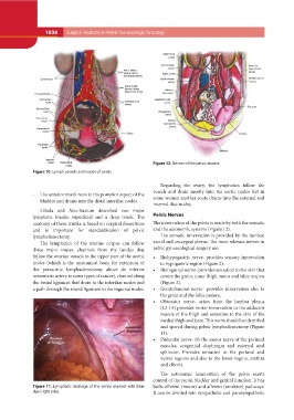
Figure 11. Lymphatic drainage of the cervix marked with blue covers the groin, inner thigh, mons and labia majora
dye (right side). (Figure 2).
• Genitofemoral nerve- provides innervation also to
the groin and the labia majora.
• Obturator nerve- arises from the lumbar plexus
(L2-L4) provides motor innervation to the adductor
muscle of the thigh and sensation to the skin of the
medial thigh and knee. This nerve should be identified
and spared during pelvic lymphadenectomy (Figure
13).
• Pudendal nerve- it’s the motor nerve of the perineal
muscles, urogenital diaphragm and external anal
sphincter. Provides sensation to the perianal and
vulvar regions and also to the lower vagina, urethra
and clitoris.
The autonomic innervation of the pelvis exerts
control of the rectal, bladder and genital function. It has
both, efferent (motor) and afferent (sensitive) pathways.
It can be divided into sympathetic and parasympathetic