
Page 16 - textbook5chapters
P. 16
Surgical Anatomy in Pelvic Gynaecologic Oncology 1037
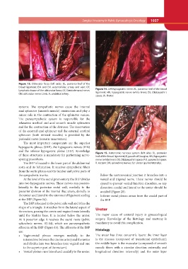
Figure 13. Obturator fossa (left side). BL, posterior leaf of the Figure 14. Left hypogastric nerve. BL, posterior leaf of the broad
broad ligament; EIA and EIV, external iliac artery and vein; OF, ligament, HN, hypogastric nerve (white lines); OS, Okabayashi´s
lymphatic tissue of the obturator fossa; GF, Genitofemoral nerve; space; Ur, Ureter.
ON, obturator nerve; Umb. A, umbilical artery.
systems. The sympathetic nerves cause the internal Figure 15. Autonomic nervous system (left side). BL, posterior
anal sphincter (smooth muscle) contraction and play a leaf of the broad ligament; D, pouch of Douglas; HN, hypogastric
minor role in the contraction of the sphincter vesicae. nerve (white lines); OS, Okabayashi´s space; PrS, pararectal space;
The parasympathetic system is responsible for the R, rectum; SN, splanchnic nerves; Ur, Ureter (pulled laterally).
relaxation urethral and anal smooth muscle sphincters
and for the contraction of the detrusor. The innervation Below the ureterovesical junction it branches into a
of the external anal sphincter and the external urethral vesical and trigonal nerve. These nerves should be
sphincter (both striated muscles) is provided by the spared to prevent vesical function disorders, so any
pudendal nerve (somatic innervation). dissection caudal and lateral to the ureter should be
avoided (Figure 20).
The most important components are the superior • Inferior rectal plexus: arises from the caudal part of
hypogastric plexus (SHP), the hypogastric nerves (HN) the IHP.
and the inferior hypogastric plexus (IHP). Knowledge
of these structures is mandatory for performing nerve- Ureter
sparing procedures.
The major cause of ureteral injury is gynaecological
The SHP is located in the lower part of the abdominal surgery. Knowledge of the histology and anatomy is
aorta and its bifurcation. It receives sympathetic fibers mandatory to avoid this complication.
from the aortic plexus and the lumbar and pelvic parts of
the sympathetic trunks. Histology
The ureter has three concentric layers: the inner layer
At the level of the sacral promontory, the SHP divides is the mucosa (composed of transitional epithelium);
into two hypogastric nerves. These nerves run postero- the middle layer is the muscular (composed of smooth
laterally to the posterior rectal wall, medially to the muscle fibers with a circular direction externally and
posterior division of the internal iliac artery, dorsally to longitudinal direction internally) and the outer layer
the ureter and lateral to the uterosacral ligament; ending
at the IHP (Figure 14).
The IHP is located in the pelvic side wall and it has the
shape of a triangle. It stretches from the lateral aspect of
the rectum, passing the cervix and vagina fornix laterally
until the bladder base. It is located below the ureter.
At it posterior edge it receives the sacral roots (pelvic
splanchnic nerves- S2-S4), which are parasympathetic
afferents of the IHP (Figure 15). The efferents of the IHP
are:
• Vaginorectal plexus: emerges medially to the
intersection between the uterine artery and the ureter
and divides into two branches (one vaginal and one
to the superior part of the rectum).
• Vesical plexus: runs lateral and caudally to the ureter.