
Page 19 - textbook5chapters
P. 19
1040 Surgical Anatomy in Pelvic Gynaecologic Oncology
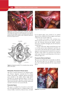
Figure 22. Parametrium and Paracervix (right side). The ureter Figure 24. Paravesical space.
runs between the superficial and the deep uterine veins (yellow
circle). DUV, deep uterine vein; IIA, internal iliac artery; SUV, su- by the superior pubic ramus, dorsally by the cardinal
perficial uterine vein; UA, uterine artery; Dotted line represents ligament and caudally by the iliococcygeus muscle (part
the superior edge of the IHP. of the levator ani muscle) (Figure 24).
Figure 23. Schematic drawing of the connective tissue and the The content of this space is the umbilical artery
spaces of the pelvis. (which separates this spaces into a medial and a lateral
part), the obturator neurovascular bundle, lymphatic
tissue and, in the lateral and upper part, the external iliac
vessels (Figure 7).
Vascular connections, either arterial (between 8 and
25 % of the cases) or venous (between 67 and 95%), can
occur between the obturator and external iliac or inferior
epigastric vessels, designated Corona Mortis or accessory
obturator vessels. These run posterioly to the superior
pubic ramus therefore careful dissection of this area is
advisable (Figure 25).
Presacral or Retrorectal Space
This space can be divided into two segments: one inferior-
the Retrorectal space and one superior the Presacral
space (that continues superiorly with the retroperitoneal
space).
Retropubic, Prevesical or Retzius Space Figure 25. Corona Mortins.
This space is limited anteriorly by the symphysis pubis
and Cooper´s ligament, posterioly by the urinary
bladder and laterally it continues with the perivesical
space (laterally to the bladder pillars) being limited
laterally by the internal obturator muscle. The floor is
the pubocervical fascia. This space contains: the dorsal
clitoral neurovascular bundle, the proximal urethra and
laterally the obturator neurovascular bundle.
Paravesical Space
This space is considered by some authors as the lateral
part of the prevesical space. It is limited medially by the
urinary bladder, laterally by obturator fascia, ventrally