
Page 20 - textbook5chapters
P. 20
Surgical Anatomy in Pelvic Gynaecologic Oncology 1041
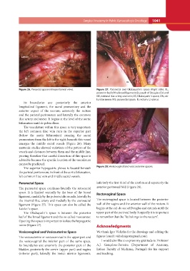
Figure 26. Presacral space (retroperitoneal view). Figure 27. Pararectal and Okabayashi´s space (Right side). BL,
posterior leaf of the broad ligament; D, pouch of Douglas; EIA and
Its boundaries are: posteriorly the anterior EIV, external iliac artery and vein; OS, Okabayashi´s space, ON, ob-
longitudinal ligament, the sacral promontory and the turator nerve; PrS; pararectal space; R, rectum; U, uterus.
anterior aspect of the sacrum; anteriorly the rectum
and the parietal peritoneum and laterally the common Figure 28. Vesicovaginal and vesicouterine spaces.
iliac artery and ureter. It begins at the level of the aortic
bifurcation until de pelvic floor. inferiorly the first third of the urethra and superiorly the
anterior peritoneal fold (Figure 28).
The vasculature within this space is very important: Rectovaginal Space
the left common iliac vein runs in the superior part The rectovaginal space is located between the posterior
(below the aortic bifurcation) crossing the sacral wall of the vagina and the anterior wall of the rectum. It
promontory from the left to the right; beneath this vessel begins at the cul-de-sac of Douglas and extends until the
emerges the middle sacral vessels (Figure 26). Many upper part of the perineal body. Surgically it is important
anatomic studies showed variations of the pattern of the to remember that the “fat belongs to the rectum”.
vessels and distances between them and the middle line,
proving therefore that careful dissection of this space is Acknowledgments
advisable because the specific location of the vasculature
cannot be predicted. We thank Igor Vidinha for the drawings and editing the
figures (email: vidinhaigor@gmail.com).
The superior hypogastric plexus is located beneath
the parietal peritoneum, in front of the aortic bifurcation, I would also like to express my gratitude to Professor
left common iliac vein and middle sacral vessels. A.J. Gonçalves-Ferreira (Department of Anatomy,
Lisbon Faculty of Medicine, Portugal) for his support
Pararectal Space and teaching.
The pararectal space continues laterally the retrorectal
space. It is limited ventrally by the base of the broad
ligament, caudally by the puborectalis muscle, laterally by
the internal iliac artery and medially by the uterosacral
ligament (Figure 25). This space can also be called the
Latzko´s space.
The Okabayashi´s space is between the posterior
leaf of the broad ligament and the so-called mesoureter.
Opening this space is important to isolate the hypogastric
nerve (Figure 27).
Vesicovaginal and Vesicouterine Space
The vesicouterine or vesicocervical is the upper part and
the vesicovaginal the inferior part of the same space.
Its boundaries are: anteriorly the posterior part of the
bladder, posteriorly the cervix (upper part) and vagina
(inferior part), laterally the vesico uterine ligaments,