
Page 13 - textbook5chapters
P. 13
1034 Surgical Anatomy in Pelvic Gynaecologic Oncology
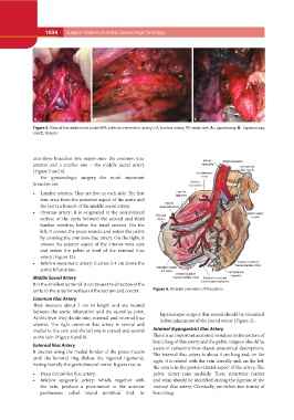
AB C
Figure 5. View of the abdominal aorta: IMA, inferior mesenteric artery; LA, lumbar artery; RV, renal vein. A. Laparotomy; B. Laparoscopy
and C. Robotic
into three branches: two major ones- the common iliac Figure 6. Arteries and veins of the pelvis.
arteries and a smaller one – the middle sacral artery
(Figure 5 and 6). laparoscopic surgery this vessels should be visualized
before placement of the lateral trocar (Figure 2).
For gynaecologic surgery the most important Internal (hypogastric) Iliac Artery
branches are: There is an important anatomic variation in the pattern of
branching of this artery and the pelvic surgeon should be
• Lumbar arteries: They are five on each side. The first aware of variations from classic anatomical descriptions.
four arise from the posterior aspect of the aorta and The internal iliac artery is about 4 cm long and, on the
the last is a branch of the middle sacral artery. right, it is related with the vein laterally and, on the left,
the vein is in the postero-lateral aspect of the artery. The
• Ovarian artery: it is originated at the anterolateral pelvic ureter runs medially. These structures (ureter
surface of the aorta between the second and third and vein) should be identified during the ligation of the
lumbar vertebra, below the renal arteries. On the internal iliac artery. Classically, we define two trunks of
left, it crosses the psoas muscle and enters the pelvis branching:
by crossing the common iliac artery. On the right, it
crosses the anterior aspect of the inferior vena cava
and enters the pelvis at level of the external iliac
artery (Figure 12).
• Inferior mesenteric artery: it arises 3-4 cm above the
aortic bifurcation.
Middle Sacral Artery
It is the smallest terminal. It continues the direction of the
aorta in the anterior surface of the sacrum and coccyx.
Common Iliac Artery
They measure about 5 cm in length and are located
between the aortic bifurcation and the sacroiliac joint.
At this level they divide into: external and internal iliac
arteries. The right common iliac artery is ventral and
medial to the vein and the left one is cranial and ventral
to the vein (Figure 6 and 8).
External Iliac Artery
It courses along the medial border of the psoas muscle
until the femoral ring (below the inguinal ligament),
having laterally the genitofemoral nerve. It gives rise to:
• Deep circumflex iliac artery.
• Inferior epigastric artery: which, together with
the vein, produce a prominence in the anterior
peritoneum called lateral umbilical fold. In