
Page 11 - textbook5chapters
P. 11
1032 Surgical Anatomy in Pelvic Gynaecologic Oncology
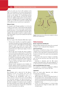
patients (it can be very short in slim patients). So the Figure 2. Vessels and nerves of the anterior abdominal wall and
angle of the introduction of the Veress needle should be location of the Palmer’s point.
close to 90º in obese patients and 45º (towards the uterine
fundus) in slim patients. The Trendelenburg position Pelvic Anatomy
changes the normal anatomy, decreasing the distance
between the umbilical region and the sacral promontory Bones, Ligaments and Muscles
(therefore increasing the risk of damage to major blood
vessels). So it should only be made after the introduction The bony pelvis is formed by the sacrum, the coccyx and
of the umbilical trocar, to avoid major complications. two hip (os coxae, innominate) bones (Figure 3).
Palmer’s Point Sacrum and Coccyx
The sacrum is formed by the fusion of the 5 sacrum
It is located in the left upper quadrant, 3 cm below the vertebrae. It has one anterior (or pelvic) and one posterior
middle left costal margin. At this point the abdominal wall (or dorsal) surface, each with four paired foramina (sacral
is relatively thin (2-3 cm) and no major retroperitoneal foramina): exit holes of the sacral nerves and anteriorly
vessels runs below it. This an alternative side for primary also the vessels.
trocar insertion for patients who have an increase risk of
umbilical adhesions but it should be avoided in patients Laterally, by the sacral alae (“wings”), it articulates
with splenomegaly and previous stomach and transverse with the hip bone (sacroiliac joint) and inferiorly with
colon surgery. the coccyx.
Blood Vessels Superiorly it articulates with the fifth lumbar
vertebrae (lumbosacral joint). The sacral promontory is
The blood supply of the abdominal wall has three origins: an anterior projection located in the first sacral vertebrae.
• Femoral artery branches: the superficial epigastric, Hip or Innominate Bones (Os Coxae)
superficial circumflex and external pudendal arteries It is formed by 3 components (originated by different
arise just below the inguinal ligament. The superficial ossification points): Ilium, Ischium and Pubis.
epigastric vessels run medial and cranially. They
can be identified during transverse incisions and by Ligaments
transillumination in laparoscopic surgery (mostly in
thin patients). There are several pelvic ligaments, with different
functions and compositions.
• External iliac artery branches: the inferior epigastric
artery and the deep circumflex artery. The first enters Surgically the most important are:
the rectus sheath at the level of the arcuate line. The • Inguinal ligament: it formed by the lower border of
inferior epigastric artery and vein can and should
be identified in laparoscopic surgery because they the aponeurosis of the external oblique muscle and it
produce the lateral umbilical fold. Theses vessels can stretches from anterior superior iliac spine to pubis. It
also be damage in transverse incisions (especially an important landmark for hernia repair and inguinal
if they go beyond the lateral limit of the rectus lymphadenectomy.
abdominis) (Figure 2). • Cooper´s or pectineal ligament: it is located along
the pectineal line, being the anterior limit of the
• Internal thoracic (mammary) artery branches: the retropubic space.
superior epigastric and the musculophrenic arteries.
The first descends in rectus sheath posterior to muscle
and anastomosis with inferior epigastric artery.
Nerves
The abdominal wall is innervated by the thoraco-
abdominal (T7-T11), the subcostal nerve (T12), the
ilioinguinal (L1) and iliohypogastric nerves (L1).
The ilioinguinal and iliohypogastric have only a
sensory function. These nerves can be injured during
low abdominal incisions and lateral placement of
laparoscopic trocars. Anatomic studies have shown that
the risk is minimized if secondary trocars are placed
above the level of the anterior superior iliac spine (Figure
2). In terms of dermatomes, it’s important to remember
that T10 corresponds to the umbilicus.