
Page 2 - sackcasestudy
P. 2
1
Introduction
Background on Takayasu’s Arteritis
Takayasu’s Arteritis (TA) is a chronic panarteritis condition of the large arteries, such as the
aorta, common carotid, and subclavian,
which causes arterial wall thickening,
stenotic changes, and potentially the
formation of aneurysmal and thrombus
development (Johnston, Lock, & Gompels,
2002; Mavrogeni, Dimitroulas,
Chatziioannou, & Kitas, 2013). TA is also
called Martorell syndrome, occlusive
thromboarteriopathy, pulseless disease,
idiopathic aortitis, aortic arch syndrome,
aortoarteritis, and stenosing aortitis
(Johnston et al., 2002; Russo & Katsicas,
2018). TA is more prevalent in women in
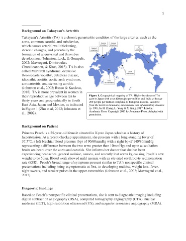
their reproductive age between ten to Figure 1. Geographical mapping of TA: Higher incidence of TA
thirty years and geographically in South seen in Japan with over 400 people per million and India with over
200 people per million compared to European nations. Adapted
East Asia, Japan and Mexico, as indicated from the heart in rheumatic, autoimmune and inflammatory diseases
in Figure 1 (Zhu et al, 2012; Johnston et (p. 390), by H. Zhang, L. Yang & X. Jiang, 2017, London:
al., 2002). Academic Press. Copyright 2017 by Academic Press. Adapted with
permission.
Background on Patient
Princess Peach is a 25-year-old female situated in Kyoto Japan who has a history of
hypertension. At a recent checkup appointment, she presents with a long-standing fever of
37.5°C; a left brachial blood pressure (bp) of 90/60mmHg with a right bp of 140/80mmHg
representing a difference between the two arms greater than 10mmHg; and upon auscultation
bruits are heard over the aorta and carotids. She informs her doctor that she has been
experiencing headaches, general malaise, nausea, and recently lost seven kg causing Peach’s new
weight to be 50kg. Blood work showed mild anemia with an elevated erythrocyte sedimentation
rate (ESR). Peach’s broad range of symptoms present similar to TA’s nonspecific clinical
presentations including being asymptomatic at first, to developing malaise, weight loss, fever,
night sweats, and weaker pulses in the upper extremities (Johnston et al., 2002; Mavrogeni et al.,
2013).
Diagnostic Findings
Based on Peach’s nonspecific clinical presentations, she is sent to diagnostic imaging including
digital subtraction angiography (DSA), computed tomography angiography (CTA), nuclear
medicine (PET), high-resolution ultrasound (US), and magnetic resonance angiography (MRA).