
Page 6 - sackcasestudy
P. 6
5
US
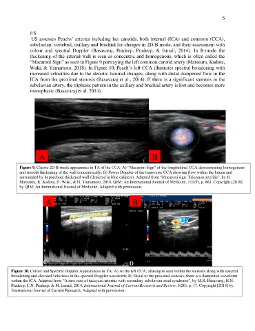
US assesses Peachs’ arteries including her carotids, both internal (ICA) and common (CCA),
subclavian, vertebral, axillary and brachial for changes in 2D-B mode, and their assessment with
colour and spectral Doppler (Basavaraj, Pradeep, Pradeep, & Ismail, 2014). In B-mode the
thickening of the arterial wall is seen as concentric and homogenous, which is often called the
“Macaroni Sign” as seen in Figure 9 portraying the left common carotid artery (Matsuura, Kadota,
Waki, & Yamamoto, 2018). In Figure 10, Peach’s left CCA illustrates spectral broadening with
increased velocities due to the stenotic luminal changes, along with distal dampened flow in the
ICA from the proximal stenosis (Basavaraj et al., 2014). If there is a significant stenosis in the
subclavian artery, the triphasic pattern in the axillary and brachial artery is lost and becomes more
monophasic (Basavaraj et al. 2014).
A B
Figure 9. Classic 2D B-mode appearance in TA of the CCA. A) “Macaroni Sign” of the longitudinal CCA demonstrating homogenous
and smooth thickening of the wall concentrically; B) Power Doppler of the transverse CCA showing flow within the lumen and
surrounded by hypoechoic thickened wall (depicted in blue calipers). Adapted from “Macaroni sign: Takayasu arteritis”, by H.
Matsuura, K. Kadota, D. Waki, & H. Yamamoto, 2018, QJM: An International Journal of Medicine, 111(9), p. 661. Copyright [2018]
by QJM: An International Journal of Medicine. Adapted with permission.
A B
Figure 10. Colour and Spectral Doppler Appearances in TA: A) At the left CCA, aliasing is seen within the stenosis along with spectral
broadening and elevated velocities in the spectral Doppler waveform; B) Distal to the proximal stenosis, there is a dampened waveform
within the ICA. Adapted from “A rare case of takayasu arteritis with secondary subclavian steal syndrome”, by M.R. Basavaraj, H.N.
Pradeep, C.N. Pradeep, & M. Ismail, 2014, International Journal of Current Research and Review, 6(20), p. 17. Copyright [2014] by
International Journal of Current Research. Adapted with permission.