
Page 8 - sackcasestudy
P. 8
7
MRA
The non-ionizing radiation modality of MRA
utilizes gadolinium-based contrast agents to
provide information on arterial vessel anatomy
(Russo & Katsicas, 2018). This includes
characteristics such as edema, thickness, and
contrast enhancement during stages of
inflammation (Russo & Katsicas, 2018).
Sensitivity and specificity of MRA detecting TA
is 100% (Mavrogenic et al., 2013). Figure 13
shows narrowing in the left subclavian artery Figure 13. MRA of a left subclavian artery stenosis. Adapted
(Mavrogeni et al., 2013). The capability of MRA from “The role in multimodality imaging in the evaluation of
to perform cross-sectional arterial wall scans Takayasu Arteritis” by S. Mavrogeni, T. Dimitroulas, S.N.
allows for detection of regions of myocardial Chatziioannou, & G. Kitas, 2013, Seminars in arthritis and
rheumatism, 42 (4), p. 409. Copyright [2013] by Seminars in
infarction, fibrosis, and intramural inflammation arthritis and rheumatism. Adapted with permission.
(Russo & Katsicas, 2018; Kissin & Merkel,
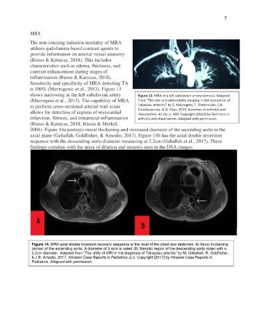
2004). Figure 14a portrays mural thickening and increased diameter of the ascending aorta in the
axial plane (Gaballah, Goldfisher, & Amodio, 2017). Figure 14b has the axial double inversion
sequence with the descending aorta diameter measuring at 2.2cm (Gaballah et al., 2017). These
findings correlate with the areas of dilation and stenosis seen in the DSA images.
Figure 14. MRA axial double inversion recovery sequence at the level of the chest and abdomen. A) Mural thickening
(arrow) of the ascending aorta. A diameter of 3.4cm is noted. B) Stenotic region of the descending aorta noted with a
2.2cm diameter. Adapted from “The utility of MRI in the diagnosis of Takayasu arteritis” by M. Gaballah, R. Goldfisher,
& J.B. Amodio, 2017, Hindawi Case Reports in Pediatrics, p.3. Copyright [2017] by Hindawi Case Reports in
Pediatrics. Adapted with permission.