
Page 3 - sackcasestudy
P. 3
2
DSA
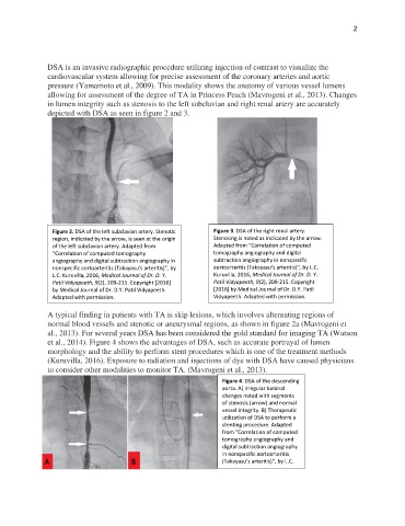
DSA is an invasive radiographic procedure utilizing injection of contrast to visualize the
cardiovascular system allowing for precise assessment of the coronary arteries and aortic
pressure (Yamamoto et al., 2009). This modality shows the anatomy of various vessel lumens
allowing for assessment of the degree of TA in Princess Peach (Mavrogeni et al., 2013). Changes
in lumen integrity such as stenosis to the left subclavian and right renal artery are accurately
depicted with DSA as seen in figure 2 and 3.
Figure 2. DSA of the left subclavian artery. Stenotic Figure 3. DSA of the right renal artery.
region, indicated by the arrow, is seen at the origin Stenosing is noted as indicated by the arrow.
of the left subclavian artery. Adapted from Adapted from “Correlation of computed
“Correlation of computed tomography tomography angiography and digital
angiography and digital subtraction angiography in subtraction angiography in nonspecific
nonspecific aortoarteritis (Takayasu’s arteritis)”, by aortoarteritis (Takayasu’s arteritis)”, by L.C.
L.C. Kuruvilla, 2016, Medical Journal of Dr. D. Y. Kuruvilla, 2016, Medical Journal of Dr. D. Y.
Patil Vidyapeeth, 9(2), 209-215. Copyright [2016] Patil Vidyapeeth, 9(2), 209-215. Copyright
by Medical Journal of Dr. D.Y. Patil Vidyapeeth. [2016] by Medical Journal of Dr. D.Y. Patil
Adapted with permission. Vidyapeeth. Adapted with permission.
A typical finding in patients with TA is skip lesions, which involves alternating regions of
normal blood vessels and stenotic or aneurysmal regions, as shown in figure 2a (Mavrogeni et
al., 2013). For several years DSA has been considered the gold standard for imaging TA (Watson
et al., 2014). Figure 4 shows the advantages of DSA, such as accurate portrayal of lumen
morphology and the ability to perform stent procedures which is one of the treatment methods
(Kuruvilla, 2016). Exposure to radiation and injections of dye with DSA have caused physicians
to consider other modalities to monitor TA. (Mavrogeni et al., 2013).
Figure 4. DSA of the descending
aorta. A) Irregular luminal
changes noted with segments
of stenosis (arrow) and normal
vessel integrity. B) Therapeutic
utilization of DSA to perform a
stenting procedure. Adapted
from “Correlation of computed
tomography angiography and
digital subtraction angiography
in nonspecific aortoarteritis
(Takayasu’s arteritis)”, by L.C.
Kuruvilla, 2016, Medical Journal
of Dr. D. Y. Patil Vidyapeeth,
9(2), 209-215. Copyright [2016]
by Medical Journal of Dr. D.Y.
Patil Vidyapeeth. Adapted with
permission.