
Page 7 - sackcasestudy
P. 7
6
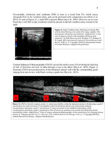
Occasionally, subclavian steal syndrome (SSS) is seen as a result from TA, which causes
retrograde flow in the vertebral artery, and can be presented with a hyperemia test (Osiro et al.
2012) As seen in Figure 11, a right SSS is present (Basavaraj et al., 2014). However, in our case
Peach has a left SSS so this waveform would be present in the left vertebral artery instead of the
right.
Figure 11. Right Vertebral artery illustrating retrograde flow
with the blood flowing to the right of the image caudally. This
demonstrates subclavian steal syndrome. Adapted from “A rare
case of takayasu arteritis with secondary subclavian steal
syndrome”, by M.R. Basavaraj, H.N. Pradeep, C.N. Pradeep, &
M. Ismail, 2014, International Journal of Current Research and
Review, 6(20), p. 17. Copyright [2014] by International Journal
of Current Research. Adapted with permission.
Contrast Enhanced-Ultrasonography (CEUS) can also be used to assess TA involving the injection
of 2mL of SonoVue and 5mL of saline through a vein in the elbow (Ma et al., 2019). Figure 12
illustrates CEUS neovascularization of the thickened arterial wall with the corresponding grade
ranging from zero to two, with Peach scoring a grade two (Ma et al., 2012).
A B C
Figure 12. CEUS of the left common carotid. A) absent microbubbles within the thickened arterial wall indicating a grade 0
classification; B) small movement of microbubbles within the thickened arterial wall (representing moderate
vascularization) indicating a grade 1 classification; C) large movement of microbubbles (representing a high
vascularization) indicating a grade 2 classification. The red arrows are pointing to the arterial wall thickening and the
microbubbles within. Adapted from “Value of contrast-enhanced ultrasonography of the carotid artery for evaluating disease
activity in “Takayasu arteritis” by L.Y. Ma et al., 2019, Arthritis Research & Therapy, 21(1), p. 3. Copyright [2019] by
Arthritis Research & Therapy. Adapted with Permission.