
Page 9 - sackcasestudy
P. 9
8
Discussion
TA is a rare disease annually affecting two per one million people worldwide with a 35 percent
mortality rate in children (Russo & Katsicas, 2018). The disease is known to manifest in three
phases. The first phase is known as the pre-pulseless period, consisting of the nonspecific
clinical presentations; the second phase is known as the vasculitic period consisting of vascular
tenderness; and the final phase is known as the late period involving stenotic symptoms
(Vaideeswar & Deshpande, 2013). The etiology is unknown for TA; however, there are genetic
factors regarding the HLA complex and infectious agents such as HIV and tuberculosis (TB)
predisposing individuals (Russo & Katsicas, 2018). The pathogenesis of TA is linked with an
immune response causing thickening of the adventitia; inflammatory cell infiltration through
the vaso vasorum leading to smooth muscle cell deterioration; and fibrosis in the intima leading
to hyperplastic proliferation and stenotic development (Russo & Katsicas, 2018; Mavrogeni et
al., 2013).
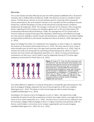
Based on findings from DSA, TA is divided into five categories, as seen in Figure 15, regarding
the location of the arterial inflammation (Zhu et al., 2012). The most common form is type V
which includes type IIb and IV and is the type Peach presents with (Zhu et al., 2012). Type IIb
involves the aortic arch along with the ascending and descending aorta, where as Type IV
involves the renal arteries and abdominal aorta (Zhu et al., 2012). Coronary and pulmonary
involvement can also occur, and in Peach’s case the diagnostic testing of CTA shows right
pulmonary involvement for Peach (Zhu et al., 2012).
Figure 15. Types of TA. Type I (far left) involving the aortic
arch branches; Type IIa involving the aortic branches, and
ascending aorta; type IIb involving the aortic branches and
ascending and descending aorta; type III involving the
descending aorta, renal and abdominal aorta; type IV
involving the renal and abdominal aorta; type V involving
type IIb and type IV. Adapted from “Takayasu arteritis:
Imaging spectrum at multidetector CT angiography” by F.P.
Zhu, S. Luo, Z.J. Wang, Z.Y Jin, L.J Zhang, & G.M. Lu,
2012, The British Journal of Radiology, 85(1020), p.1282.
Copyright [2012] by the British Journal of Radiology.
Adapted with permission.
TA is often difficult to diagnose in a clinical setting due to nonspecific signs and symptoms, the
lack of serological findings, along with the lack of clinical expertise of this rare condition
(Mavrogeni et al., 2013). This leads to a late clinical diagnosis where arterial damage is
established (Mavrogeni et al., 2013).
According to the Sharma criteria for diagnosis, as seen in Table 1, the patient has a high
probability of having TA with the presentation of either two major criteria, one major with two
minor or four minor criteria (Zhu et al., 2012). Sharma criteria is preferred over Ishikawa’s and
the American college of Rheumatology as there is a higher sensitivity and specificity with the
former, and the latter criteria lacks recent revision regarding the non-invasive imaging
modalities (Mavrogeni et al., 2013; Zhu et al., 2012).