Page 209 - 00. Complete Version - Progress Report IPEN 2014-2016
P. 209
Nuclear Science and Technology | Progress Report  209


               dosimetry, in order to assess the viability of   diodiagnostic images and quality assurance
               building a phantom for radiotherapy with a     of dosimetry in radiation therapy area. The
               3D printer. The results indicate that the PLA   development of simulators using 3D print-
               printed plates can be considered tissue-equiv-  ers and tissue-equivalent materials proved
               alent and agree with the obtained using sol-   to be quite feasible for use in the field of vet-
               id water plates.                               erinary radiology. The images obtained by CT
                                                              exams showed good agreement with real ex-
               Development of tissue equivalent phantoms      ams with animals, thus this dog’s phantom
               using 3D printer for application in quality    has demonstrated to be very useful for quality
               assurance in veterinary medicine               control in radiodiagnostic and radiation thera-

                                                              py areas. The methodology used in this study
                                                              enabled the development of personalized ex-
                                                              ams for the verification of a better protocol on
                                                              the evaluation of absorbed doses in patients.
                                                              This work will allow the creation and devel-
                                                              opment of phantom of different shapes and
                                                              sizes with tissue-equivalent material that will
                                                              be used for application in veterinary medicine
                                                              (radiodiagnosis and radiation therapy). Parts
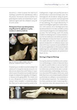
                                                              of the printed dog phantom are presented in
                                                              Figure 10, 11.


                                                              Metrology in Diagnostic Radiology


                                                              During the period of 2014-2016, a new calibra-
               Figure 10 - Printed parts: (a)Brain; (b) Skull – lateral view;
               (c) Dog head – external view; (d) Skull – frontal view.  tion methodology in terms of absorbed dose
                                                              to water for parallel plate ionization chambers
               3D printing is a revolutionary technology that   to be used in low energy X rays beams (10kV
               allows us to change the way we consume, cre-   to 100kV) was established. In order to estab-
               ate and even live in the world. Today(,) it is pos-  lish this calibration methodology, two ioniza-
               sible to print 3D anatomical models based on   tion chambers PTW models 23344 were used.
               images of computed tomography (CT), Magnet-    Both chambers were characterized and tested
               ic Resonance Imaging (MRI). This work aims     qualitatively according to recommendations
               to use a 3D printer to build a dog phantom     of international standards. The characteriza-
               named “canis morphic”, a veterinary phan-      tion tests performed were: measuring effec-
               tom that can be used in quality control of ra-  tive minimum dose rate measurements, the


               Figure 11- Printed parts: (a) cervical spine; (b) lumbar spine.

