
Page 109 - Science
P. 109
RESEARCH | REPORT
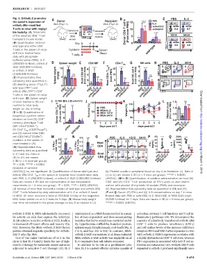
Fig. 3. OrthoIL-2 promotes PBS IL-2
the specific expansion of Donor Recipient 15 **** PBS
IL-2
orthoIL-2Rb–modified BL6 (Thy1.1) BL6 (Thy1.2) ortho1G12
T cells in mice with negligi- 10 ortho3A10
Transduce Transplant
ble toxicity. (A) Schematic CD8 CD8 Donor CD8 T cell # (Fold over PBS) ****
+
of the adoptive CD8 T cell CD8 mixture 5 ns 1G12 3A10
transplant mouse model. enrich YFP * ns
(B) Quantification of donor Day: 0 4 5 0
wild-type and ortho CD8 + ortho Thy1.1
T cells in the spleen of recip- Cytokine: Spleen wild-type YFP
ient mice treated twice T cells: Blood
daily with phosphate-
buffered saline (PBS), IL-2
(250,000 IU/dose), orthoIL-2 Spleen weight CD8 MP CD4 T reg NK cells PBS IL-2 3A10
1G12 (250,000 IU/dose), 10 **** **** 15 **** 4 ****
or orthoIL-2 3A10 6 3 NK1.1
(2,500,000 IU/dose). Spleen weight (mg/g) ns Host CD8 MP (Fold over PBS) 4 Host CD4+ T reg # (over PBS) 10 Host NK cell # (Fold over PBS) ns
(C) Representative flow 5 ns ns 2 CD49b
cytometry data quantified in 2 ns 5 ns 1
+
(B) depicting donor (Thy1.1 ) 0 0 0 0 Foxp3
–
ortho3A10
ortho3A10
ortho3A10
wild-type (YFP ) and PBS IL-2 PBS IL-2 PBS IL-2 PBS IL-2
ortho3A10
+
orthoIL-2Rb (YFP )CD8 + ortho1G12 ortho1G12 ortho1G12 ortho1G12 CD25
T cells in the spleen of recip-
ient mice. (D) Spleen weight
of mice treated in (B) nor- PBS IL-2 ortho1G12 100 1.5 ns Downloaded from
malized to total body 15 *** 110 ns
weight on day of killing. Survival (%) PBS Weight (%) Platelet count (Fold over PBS) 1.0
MSA-IL-2
(E to G) Quantification of Donor CD4 T eff # (Fold over PBS) 10 50 MSA-1G12 100 0.5 ****
exogenous cytokine admin- MSA-3A10 90 ****
istration on host (E) CD8 + 5 * * * 0 0.0
MSA-3A10
memory phenotype T cell 0 0 1 2 3 4 5 0 1 2 3 4 5 PBS IL-2
+
+
(MP, CD44 CD62L ), ortho Days post ACT Days post ACT MSA-1G12
+
+
+
(F) CD4 T reg (CD25 Foxp3 ), wild-type
and (G) natural killer (NK) http://science.sciencemag.org/
+
+
cell (CD3-NK1.1 CD49b ) 2.0 **** PBS IL-2 1G12 3A10 25 **** 3000 ****
numbers in the spleen of 20 **** 20 2000
+ + 1.5 1000
mice treated in (A). 15 ns Host IFN (pg/mL) 15 IL-5 (pg/mL) 10 ns
(H) Representative flow % IFN CD8 T cells 10 ns % IFN CD4 T cells 1.0 CD4 10 5 ns
cytometry data as quantified 5 0.5 5 ns ns 0
in (F) and (G). Data in 0 0.0 Host 0 PBS PBS
MSA-1G12
MSA-1G12
ortho3A10
ortho3A10
(B) to (H) are means PBS IL-2 PBS IL-2 CD8 IFN MSA-IL-2 MSA-3A10 MSA-IL-2 MSA-3A10
±SD (n = 5 mice per group). ortho1G12 ortho1G12 IL-2 on March 1, 2018
*P < 0.05, ****P < 0.0001
[analysis of variance
(ANOVA)]; ns, not significant. (I) Quantification of donor wild-type and (L) Platelet counts in peripheral blood on day 4 as treated in (J). Data in
+
orthoIL-2Rb CD4 T eff in the spleen of recipient mice treated once daily (J) to (L) are means ± SD (n = 5 mice per group). ****P < 0.0001
with PBS, IL-2 (250,000 IU/dose), or orthoIL-2 1G12 (1,000,000 IU/dose). (ANOVA). (M to O) Quantification of cytokine administration on host (M)
+
+
Data are means ± SD and are representative of two independent CD8 and (N) CD4 Tcell production of IFN-g upon ex vivo restim-
experiments (n = 4 mice per group). *P < 0.05, ***P < 0.001 (ANOVA). ulation with phorbol 12-myristate 13-acetate (PMA) and ionomycin.
(J) Survival of mice that received a mixture of wild-type and orthoIL-2Rb (O) Representative flow cytometry data as quantified in (M) and (N).
+
CD8 T cells followed by daily administration of IL-2 or orthoIL-2 fused (P and Q) Serum (P) IFN-g and (Q) IL-5 concentrations on day 7 in mice
to MSA. All mice received a total of 250,000 IU/day of the respective treated daily with PBS or with MSA–IL-2, MSA-1G12, or MSA-3A10 (each
MSA fusion protein on an IL-2 basis for 5 days. (K) Mouse body weight 25,000 IU/dose) for 7 days. Data are means ± SD (n = 5 mice per group).
over time normalized to the group average on day 0 as treated in (J). ****P < 0.0001 (ANOVA).
orthoIL-2 1G12 to MSA substantially increased administered as a MSA fusion resulted in a num- activating cytotoxic T cell functions and T cell in-
its activity on cells that express the wild-type ber of dose-dependent and dose-accumulating flammatory pathways (19). We determined the
IL-2R relative to native orthoIL-2 1G12, leading toxicities that led to weight loss, restricted mobil- capacity of adoptively transferred orthoIL-2Rb
+
to increased off-target effects and toxicity (fig. ity, hypothermia, ruffled fur, hunched posture, CD8 T cells to produce interferon-g (IFN-g)
S15). However, the MSA–orthoIL-2 3A10 fusion splenomegaly, lymphomegaly, and death (Fig. 3, and cell surface levels of the immune inhibitory
protein retained exquisite specificity for orthoIL- J to L, and figs. S15 to S18). In contrast, MSA– receptors PD-1 and TIM-3 after expansion in vivo
2Rb T cells (fig. S16). orthoIL-2 3A10 wasnontoxicat all dosesevaluated. with orthoIL-2. TIM-3 expression correlates with
+
One of the major limitations of IL-2 in the MSA–orthoIL-2 3A10 activity was negligible on all ahighlydysfunctional CD8 T cell state, whereas
clinic is that IL-2 toxicity limits the use of high- IL-2–responsive host cell subsets evaluated. PD-1 expression is associated with both T cell ac-
dose IL-2 therapy for metastatic cancer and as an In addition to itsroleasa proliferative cyto- tivation and exhaustion (20). OrthoIL-2Rb T cells
adjuvant to adoptive T cell therapy (12). IL-2 kine, IL-2 is a potent effector cytokine capable of expanded in orthoIL-2 produced significantly more
Sockolosky et al., Science 359, 1037–1042 (2018) 2 March 2018 4of6