
Page 114 - Science
P. 114
RESEARCH | REPORT
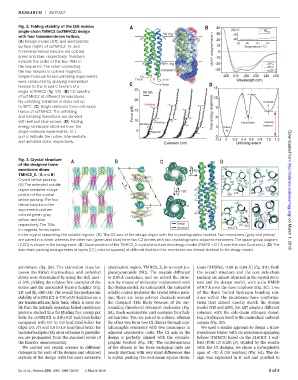
Fig. 2. Folding stability of the 156-residue
single-chain TMHC2 (scTMHC2) design
with four transmembrane helices.
(A) Design model (left) and electrostatic
surface (right) of scTMHC2. N- and
C-terminal helical hairpins are colored
green and blue, respectively. Numbers
indicate the order of the four TMs in
the sequence. The linker connecting
the two hairpins is colored magenta.
Single-molecule forced unfolding experiments
were conducted by applying mechanical
tension to the N and C termini of a
single scTMHC2 (fig. S5). (B) CD spectra
of scTMHC2 at different temperatures.
No unfolding transition is observed up
to 95°C. (C) Single-molecule force-extension
traces of scTMHC2. The unfolding
and refolding transitions are denoted
with red and blue arrows. (D) Folding
energy landscape obtained from the
single-molecule experiments. N, I,
and U indicate the native, intermediate,
and unfolded state, respectively. Downloaded from
Fig. 3. Crystal structure
of the designed trans-
membrane dimer
TMHC2_E. (A and B)
Crystal lattice packing.
(A) The extended soluble
region mediates a large http://science.sciencemag.org/
portion of the crystal
lattice packing.The four
helical hairpins in the
asymmetric unit are
colored green, gray,
yellow, and blue,
respectively.The TMs,
in magenta, forms layers
in the crystal separating the soluble regions. (B) The C2 axis of the design aligns with the crystallographic twofold. Two monomers (gray and yellow) on March 1, 2018
are paired in a dimer, whereas the other two (green and blue) form two C2 dimers with two crystallographic adjacent monomers.The space group diagram
(C121) is shown in the background. (C) Superposition of the TMHC2_E crystal structure and design model (RMSD = 0.7 Å over the core Ca atoms). (D) The
side-chain packing arrangements at layers [(C), colored squares] at different depths in the membrane are almost identical to the design model.
prominent (fig. S9). The transition rates be- cytoplasmic region, TMHC2_E, in n-nonyl-b-D- tions (RMSDs), 0.60 to 0.84 Å] (fig. S11). Both
tween the folded, intermediate, and unfolded glucopyranoside (NG). The crystals diffracted the overall structure and the core side-chain
states were determined by using the Bell mod- to 2.95-Å resolution, and we solved the struc- packing are almost identical in the crystal struc-
el (16), yielding the relative free energies of the ture by means of molecular replacement with ture and the design model, with a Ca RMSD
states and the associated barrier heights (Fig. the design model. As anticipated, the extended of 0.7 Å over the core residues (Fig. 3C). Two
2D and fig. S10) (14). The overall thermodynamic soluble region mediates the crystal lattice pack- of the three buried hydrogen bonding resi-
stability of scTMHC2 is 7.8(±0.9) kcal/mol on a ing; there are large solvent channels around dues within the membrane have conforma-
per transmembrane helix basis, which is more sta- the designed TMs likely because of the sur- tions that almost exactly match the design
ble than the naturally occurring helical membrane rounding disordered detergent molecules (Fig. model (S13 and Q93), but Q17 adopts a different
proteins studied thus far [folding free energy per 3A). Each asymmetric unit contains four heli- rotamer, with the side-chain nitrogen donat-
helixfor scTMHC2 is2.0(±0.2) kcal/(molhelix) cal hairpins: Two are paired in a dimer, whereas ing a hydrogen bond to the main-chain carbonyl
compared with 0.7 to 0.9 kcal/(molhelix) for the other two form two C2 dimers through crys- oxygen (Fig. 3D).
GlpG (14, 17) and 1.6 to 1.8 kcal/(molhelix) for tallographic symmetry with two monomers in We used a similar approach to design a trans-
bacteriorhodopsin (18); error estimates in parenthe- adjacent asymmetric units. The C2 axis in the membrane trimer with six membrane-spanning
ses are propagated from the standard errors of design is perfectly aligned with the crystallo- helices (TMHC3) based on the 5L6HC3_1 scaf-
the kinetics measurements]. graphic twofold (Fig. 3B). The conformations fold (PDB ID 5IZS) (8). Guided by the results
We carried out crystal screens in different of the dimers in the three biological units are with the C2 designs, we chose a hydrophobic
detergents for each of the designs and obtained nearly identical, with very small differences due span of ~30 Å (20 residues) (Fig. 4A). The de-
crystals of the design with the most extensive to crystal packing [Ca root-mean-square devia- sign was expressed in E. coli and purified to
Lu et al., Science 359, 1042–1046 (2018) 2 March 2018 3of 5