
Page 2552 - Cote clinical veterinary advisor dogs and cats 4th
P. 2552
1284 Syncope Tachycardia
(Continued from previous page)
VetBooks.ir Differential Diagnosis Key Features
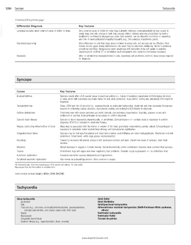
Laryngeal paralysis (most common cause of stridor in dogs)
Very common cause of stridor in older dogs (Labrador retrievers overrepresented) but also occurs in
young dogs and cats; change in bark may precede stridor; distress episodes precipitated by heat or
excitement; confirmed by laryngoscopy under light sedation; can be idiopathic (common) or caused by
any form of neuropathy/junctionopathy/myopathy (e.g., tick paralysis, myasthenia gravis)
Nasopharyngeal polyp More often seen in cats than dogs; more common in young cats, but any age can be affected; often
follows chronic upper airway inflammation; otic exam may be abnormal unilaterally; Horner’s syndrome
sometimes identified; laryngoscopy under anesthesia with retraction of the soft palate to examine
nasopharynx to confirm; CT or sometimes skull radiographs also useful but not always necessary
Neoplasia Intra- or extraluminal neoplasia/tumor; in cats, squamous cell carcinoma common; tissue biopsy required
for diagnosis
Syncope
Causes Key Features
Bradyarrhythmias Syncope results after >6-8 second pause in electrical activity (i.e., failure of subsidiary pacemaker in third-degree AV block,
or sinus arrest with subsidiary pacemaker failure in sick sinus syndrome). Auscultation, resting and ambulatory ECG helpful in
diagnosis.
Tachyarrhythmias Rates >300 bpm for >6 seconds (i.e., supraventricular or ventricular tachycardia). Heart rate and time necessary for syncope
depend on underlying cardiac structure. Auscultation, resting and ambulatory ECG helpful in diagnosis.
Outflow obstructions Commonly seen with severe pulmonic and aortic stenosis and pulmonary hypertension. Typically, syncope occurs with
excitement or exertion. Echocardiogram is necessary to confirm diagnosis.
Cyanotic heart disease Syncope is due to hypoxemia, hyperviscosity, or arrhythmia. Echocardiogram +/− contrast study is necessary to confirm
diagnosis. PCV/TS is indicated to determine therapy.
Masses obstructing inflow/outflow of blood Mass may be associated with the interior or exterior of the heart, potentially compromising cardiac output. Echocardiogram is
necessary to determine tumor location/likely etiology and hemodynamic significance.
Congestive heart failure Syncope may be first sign that animal is in heart failure before overt infiltrates are noted radiographically. Mechanism not well
understood. Small-breed, white dogs appear overrepresented.
Neurologic Caused by increased intracranial pressure with decreased cerebral perfusion. Uncommon cause of syncope, more likely
seizures.
Metabolic Abrupt decrease in oxygen or nutrient delivery. Serum biochemistry profile is indicated. Seizures more common than syncope.
Tussive Small-breed dogs with upper and lower respiratory tract problems. Consider cough suppressant +/− bronchodilator trial.
Autonomic dysfunction Excessive baroreflex causing bradycardia and hypotension.
Peripheral vasomotor dysfunction Also known as postural hypotension. More common in people.
AV, Atrioventricular; ECG, electrocardiogram; PCV, packed cell volume; TS, total solids.
Reproduced from the third edition in modified form.
THIRD EDITION AUTHOR: Sarah J. Miller, DVM, DACVIM
Tachycardia
Sinus tachycardia Atrial flutter
Excitement Atrial fibrillation
Pain Atrial/junctional tachycardias
Toxicosis (e.g., chocolate; aminophylline/theophylline; pseudoephedrine, Atrioventricular reentrant tachycardias (Wolff-Parkinson-White syndrome,
phenylpropanolamine, and related compounds; Bufo toad) others)
Sepsis Ventricular tachycardia
Hypotension Ventricular flutter
Congestive heart failure Torsade de pointes
Systemic illness (e.g., hyperthyroidism, fever, anemia)
.ExpertConsult.com
www
www.ExpertConsult.com