
Page 1053 - Small Animal Internal Medicine, 6th Edition
P. 1053
CHAPTER 57 Neonatology and Pediatrics 1025
of caloric restriction, physical therapy, and improved trac- ultrasound (Fig. 57.25, A, 57.25, B). Renal dysplasia is a
tion in the nest box. Placement of loose hobbles helps control heritable problem in several canine breeds; ultrasound can
VetBooks.ir limb movements and promotes normal ambulation within identify the typical marked morphologic abnormalities in
puppies at 6 to 8 weeks of age in breeds at risk (Fig. 57.26)
days (Fig. 57.24, B, 57.24, C, 57.24, D). The prognosis for
Congenital renal polycystic disease of brachycephalic cats
swimmer puppies treated before 4 to 5 weeks of age is good.
can similarly be identified with ultrasound in 8- to 12-week-
Urogenital Disorders old kittens (Fig. 57.27). Until reliable genetic markers are
Dysuria, urinary incontinence, or hematuria/pyuria can available for the various breed-specific congenital renal dys-
accompany neonatal urogenital disorders. The presence of a plasias, ultrasound provides the best method of screening
persistent urachus causes micturition through the umbilicus. young dogs and cats for these disorders; clinical signs are
Urachal diverticula can predispose the bladder to recurrent usually not present until early adulthood when uremia
infection because of abnormal urine flow in the region; sur- develops. Neonatal urolithiasis, with or without associated
gical excision is indicated. The diagnosis is confirmed with urinary tract infection, can cause outflow obstruction and
A B
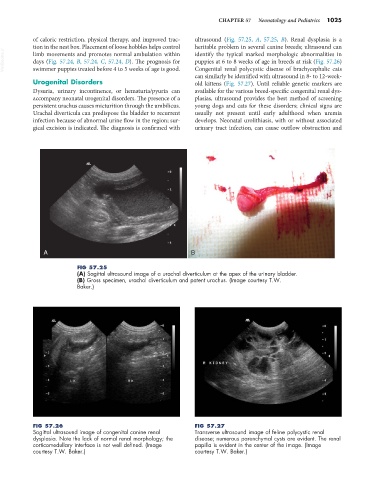
FIG 57.25
(A) Sagittal ultrasound image of a urachal diverticulum at the apex of the urinary bladder.
(B) Gross specimen, urachal diverticulum and patent urachus. (Image courtesy T.W.
Baker.)
FIG 57.26 FIG 57.27
Sagittal ultrasound image of congenital canine renal Transverse ultrasound image of feline polycystic renal
dysplasia. Note the lack of normal renal morphology; the disease; numerous parenchymal cysts are evident. The renal
corticomedullary interface is not well defined. (Image papilla is evident in the center of the image. (Image
courtesy T.W. Baker.) courtesy T.W. Baker.)