
Page 1057 - Small Animal Internal Medicine, 6th Edition
P. 1057
CHAPTER 57 Neonatology and Pediatrics 1029
VetBooks.ir
A
B C
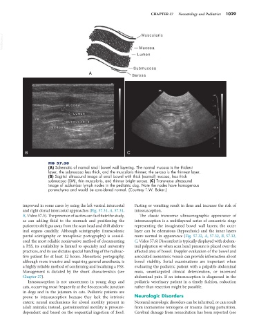
FIG 57.30
(A) Schematic of normal small bowel wall layering. The normal mucosa is the thickest
layer, the submucosa less thick, and the muscularis thinner; the serosa is the thinnest layer.
(B) Sagittal ultrasound image of small bowel with thick (normal) mucosa, less thick
submucosa (SM), thin muscularis, and thinner bright serosa. (C) Transverse ultrasound
image of sublumbar lymph nodes in the pediatric dog. Note the nodes have homogenous
parenchyma and would be considered normal. (Courtesy T.W. Baker.)
improved in some cases by using the left ventral intercostal Fasting or vomiting result in ileus and increase the risk of
and right dorsal intercostal approaches (Fig. 57.31, A, 57.31, intussusception.
B, Video 57.5). The presence of ascites can facilitate the study, The classic transverse ultrasonographic appearance of
as can adding fluid to the stomach and positioning the intussusception is a multilayered series of concentric rings
patient to shift gas away from the scan head and shift abdom- representing the invaginated bowel wall layers; the outer
inal organs caudally. Although scintigraphy (transcolonic layer can be edematous (hypoechoic) and the inner layers
portal scintigraphy or transplenic portography) is consid- more normal in appearance (Fig. 57.32, A, 57.32, B, 57.32,
ered the most reliable noninvasive method of documenting C, Video 57.6) Discomfort is typically displayed with abdom-
a PSS, its availability is limited to specialty and university inal palpation or when scan head pressure is placed over the
practices, and its use dictates special handling of the radioac- affected area of bowel. Doppler evaluation of the bowel and
tive patient for at least 12 hours. Mesenteric portography, associated mesenteric vessels can provide information about
although more invasive and requiring general anesthesia, is bowel viability. Serial examinations are important when
a highly reliable method of confirming and localizing a PSS. evaluating the pediatric patient with a palpable abdominal
Management is dictated by the shunt characteristics (see mass, unanticipated clinical deterioration, or increased
Chapter 27). abdominal pain. If an intussusception is diagnosed in the
Intussusception is not uncommon in young dogs and pediatric veterinary patient in a timely fashion, reduction
cats, occurring most frequently at the ileocecocolic junction rather than resection might be possible.
in dogs and in the jejunum in cats. Pediatric patients are
prone to intussusception because they lack the intrinsic Neurologic Disorders
enteric neural mechanisms for aboral motility present in Neonatal neurologic disorders can be inherited, or can result
adult animals; instead, gastrointestinal motility is pressure- from intrauterine teratogens or trauma during parturition.
dependent and based on the sequential ingestion of food. Cerebral damage from resuscitation has been reported (see