
Page 1058 - Small Animal Internal Medicine, 6th Edition
P. 1058
VetBooks.ir
A
B C
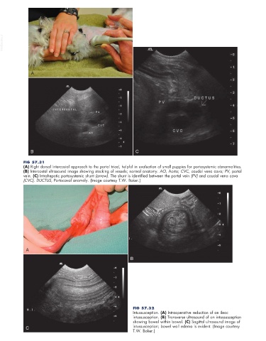
FIG 57.31
(A) Right dorsal intercostal approach to the portal triad, helpful in evaluation of small puppies for portosystemic abnormalities.
(B) Intercostal ultrasound image showing stacking of vessels; normal anatomy. AO, Aorta; CVC, caudal vena cava; PV, portal
vein. (C) Intrahepatic portosystemic shunt (arrow). The shunt is identified between the portal vein (PV) and caudal vena cava
(CVC). DUCTUS, Portocaval anomaly. (Image courtesy T.W. Baker.)
A
B
FIG 57.32
Intussusception. (A) Intraoperative reduction of an ileac
intussusception. (B) Transverse ultrasound of an intussusception
showing bowel within bowel. (C) Sagittal ultrasound image of
C intussusception; bowel wall edema is evident. (Image courtesy
T.W. Baker.)