
Page 1054 - Small Animal Internal Medicine, 6th Edition
P. 1054
1026 PART VIII Reproductive System Disorders
signs of acute abdominal pain. Lower urinary tract infection ultrasonography, cystoscopy, helical computed tomography,
in neonates has the potential for ascending and causing or a combination of these diagnostic procedures. Other con-
VetBooks.ir pyelonephritis if not detected and controlled, making genital abnormalities can also occur in dogs with ectopic
ureters, including but not limited to renal agenesis or dys-
ultrasound-guided cystocentesis preferable when acquiring
urine from pediatric patients with signs of lower tract disease.
Ectopic ureters can cause incontinence during the post- plasia, and hydronephrosis and/or hydroureter; therefore,
it is essential to evaluate the entire urinary system using
partum period; clinical signs are most evident after weaning ultrasonography if cystoscopy is the only other diagnostic
when the dam is no longer cleaning the puppy. An ectopic utilized before surgery. Accompanying urethral sphinc-
ureter is defined as one which distally enters the genitouri- ter mechanism anomalies are commonly present in dogs
nary tract in any location other than the trigone of the with ectopia, making the prognosis for complete resolu-
bladder. Ectopic ureters are the most common cause of tion of incontinence after resolution of the ectopic ureter
urinary tract infection in young dogs. Breeds reported to be guarded. Visualization of a nonvascular fluid-filled struc-
at risk include the Golden Retriever, Labrador Retriever, ture with a hyperechoic wall passing dorsal to the urinary
Siberian Husky, Newfoundland, and English Bulldog. The bladder or obvious insertion of the structure into the proxi-
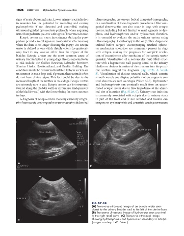
condition should be considered heritable. Ectopic ureters are mal urethra suggest the diagnosis (Fig. 57.28, A, 57.28,
uncommon in male dogs and, if present, these animals often B). Visualization of distinct ureteral walls, which contain
do not have clinical signs. This fact could be due to the smooth muscle and display pulsatile motion, supports ure-
increased length of the urethra in male dogs. Ectopic ureters teral abnormalcy such as ectopia (Video 57.3). Hydroureter
are extremely rare in cats. Ectopic ureters can be intramural and hydronephrosis can eventually result from an uncor-
(tunnel along the bladder wall) or extramural (independent rected ectopic ureter due to flow impedance at the abnor-
of the bladder wall) with the former being far more common mal site of insertion (Fig. 57.28, C). Urinary tract infection
in dogs. is commonly associated with ectopia due to urinary stasis
A diagnosis of ectopia can be made by excretory urogra- in part of the tract and, if not detected and treated, can
phy, fluoroscopic urethrography or ureterography, abdominal progress to pyelonephritis and ureteritis causing permanent
A
B
FIG 57.28
(A) Transverse ultrasound image of an ectopic ureter seen
dorsal to the urinary bladder and to the left of the uterine horn.
(B) Transverse ultrasound image of hydroureter seen proximal
to the right renal pelvis. (C) Transverse ultrasound image
C showing hydronephrosis and hydroureter secondary to ectopia.
(Images courtesy T.W. Baker.)