
Page 1118 - Small Animal Internal Medicine, 6th Edition
P. 1118
1090 PART IX Nervous System and Neuromuscular Disorders
VetBooks.ir
Hypothalamus
Brainstem
Retrobulbar
region
Middle
ear
cavity
Spinal
cord
Cranial
cervical
ganglion
Cervical thoracic T1-T4
sympathetic trunk spinal cord A
(neck) segments
Cervical thoracic
sympathetic trunk Ventral
(cranial mediastinum roots
and thoracic inlet) T1-T4
FIG 61.7
Sympathetic innervation to the eye. An injury anywhere
along this pathway will result in Horner syndrome.
the thoracic spinal cord. Upper motor neuron lesions in the
brainstem or cervical spinal cord are a relatively rare cause B
of Horner syndrome but may occur secondary to trauma,
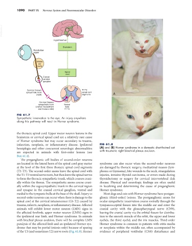
infarction, neoplasia, or inflammatory disease. Ipsilateral FIG 61.8
hemiplegia and other concurrent neurologic abnormalities (A) and (B) Horner syndrome in a domestic short-haired cat
are expected in animals with first-order lesions (see with traumatic right brachial plexus avulsion.
Box 61.4).
The preganglionic cell bodies of second-order neurons
are located in the lateral horn of the spinal cord gray matter syndrome can also occur when the second-order neurons
at the level of the first three thoracic spinal cord segments are damaged by thoracic surgery, mediastinal masses (lym-
(T1-T3). The second-order axons leave the spinal cord with phoma or thymoma), bite wounds to the neck, strangulation
the T1-T3 ventral nerve roots, but then leave the spinal nerves injuries, invasive thyroid carcinoma, or errors made during
to form the thoracic sympathetic trunk, which courses crani- thyroidectomy or surgery for cervical intervertebral disk
ally within the thorax. The sympathetic axons course crani- disease. Physical and neurologic findings are often useful
ally within the vagosympathetic trunk in the cervical region in localizing and determining the cause of preganglionic
and synapse in the cranial cervical ganglion, ventral and Horner syndrome.
medial to the tympanic bulla at the base of the skull. Injury to Most dogs and cats with Horner syndrome have postgan-
second-order neurons can occur when there is damage to the glionic (third-order) lesions. The postganglionic axons for
spinal cord at the cervical intumescence (C6-T2) caused by ocular sympathetic innervation course rostrally through the
trauma, infarcts, neoplasia, or inflammatory disease. Affected tympanooccipital fissure into the middle ear and enter the
animals will exhibit lower motor neuron (LMN) signs in cranial cavity with the glossopharyngeal nerve (CN9),
the affected forelimb, upper motor neuron (UMN) signs in leaving the cranial cavity via the orbital fissure for distribu-
the ipsilateral rear limb, and Horner syndrome. In animals tion to the smooth muscle of the orbit, the upper and lower
with brachial plexus avulsion, there will be complete LMN eyelids, the third eyelid, and the iris muscles. Third-order
paralysis of the affected limb and an ipsilateral Horner syn- Horner syndrome is common in patients with otitis media
drome that may be partial (miosis only) because of sparing or neoplasia within the middle ear, often accompanied by
of the T3 (and sometimes T2) nerve roots (Fig. 61.8). Horner evidence of peripheral vestibular (CN8) disturbance and