
Page 1119 - Small Animal Internal Medicine, 6th Edition
P. 1119
CHAPTER 61 Loss of Vision and Pupillary Abnormalities 1091
neurologic, and otoscopic examinations. Neurologic find-
ings can often help to locate the site of disruption of sympa-
VetBooks.ir thetic innervation. Further tests should be recommended
after lesion localization. Careful palpation of the soft tissues
of the neck and radiographs of the thorax and the cervical
and thoracic spine should be performed and advanced diag-
nostic imaging (MRI) should be considered if a first- or
second-order lesion is suspected. When a postganglionic
lesion is suspected, skull radiographs, CT, or MRI should be
performed to evaluate the middle ear for signs of otitis
media, neoplasia, or trauma. The most common causes of
Horner syndrome in cats are middle ear disease and injury
to the soft tissues of the neck. Dogs more frequently have
brachial plexus avulsion, nerve root tumors of the brachial
plexus, C6-T2 spinal cord injury, middle ear disease, and
cervical soft tissue injuries or tumors. Most dogs with Horner
syndrome (>50%) have no other neurologic abnormalities,
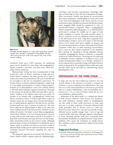
FIG 61.9 and a cause for their Horner syndrome is not identified;
Dramatic muscle atrophy in a dog with masticatory muscle these animals are classified as having idiopathic disease.
myositis has resulted in retraction of the globes into the Idiopathic postganglionic Horner syndrome is especially
orbits and protrusion of the third eyelid over most of the
corneal surface. common in Golden Retrievers. The prognosis for resolution
of idiopathic Horner syndrome is excellent as most cases
resolve spontaneously within 4 to 6 months. Treatment is
sometimes facial nerve (CN7) paralysis. No underlying rarely required, but occasionally in dogs with bilateral disease
cause can be identified in most dogs with postganglionic vision is obscured by the prolapsed third eyelids and some
Horner syndrome, and these cases have been called idio- periodic relief can be provided by administering topical
pathic Horner syndrome. phenylephrine (1%).
Pharmacologic testing has been recommended to help
localize the cause of Horner syndrome in dogs and cats.
When Horner syndrome has been present for at least 2 PROTRUSION OF THE THIRD EYELID
weeks, denervation hypersensitivity will occur in the affected
eye secondary to the loss of sympathetic innervation. To In dogs and cats the third eyelid may protrude over the
localize Horner syndrome pharmacologically, a single drop corneal surface in the presence of corneal or conjunctival
of a very dilute concentration of a direct-acting sympatho- irritation, or space-occupying retroorbital disease. This may
mimetic (0.1% phenylephrine: stock 10% solution diluted also occur if the animal experiences a decrease in periorbital
1 : 100 with saline solution) is applied to both eyes. The pupil mass as a result of dehydration, a loss of retrobulbar fat or
in the normal eye should not dilate with that dilute concen- muscle (Fig. 61.9), or a loss of volume within the eye (i.e.,
tration. Dilation of the affected pupil will occur within 20 microphthalmos, phthisis bulbi).
minutes in an animal with a postganglionic (third-order Protrusion of the third eyelid is a conspicuous feature
Horner syndrome) lesion. If neither pupil dilates within 20 of both Horner syndrome (with miosis) and dysautono-
minutes, repeat the test using a more concentrated adrener- mia (with mydriasis). Systemic illness or tranquilization
gic (1% phenylephrine) applied to the affected eye and then can also result in third eyelid protrusion in some dogs
monitor pupil size every 2 minutes in both eyes. The time and cats. A peculiar syndrome of dramatic bilateral third
that it takes for the affected pupil to dilate to the same size eyelid protrusion with no obvious cause (i.e., Haw syn-
as the unaffected pupil is the test endpoint. If dilation occurs drome) has been observed in cats and occasionally in dogs.
within 20 minutes, the lesion is most likely postganglionic Affected cats are usually younger than 2 years of age and
(third order). Dilation in 20 to 45 minutes has been reported in good health otherwise, although digestive disturbances
in dogs with second-order lesions and 60 to 90 minutes in or heavy intestinal parasite loads have occasionally been
dogs with central lesions and dogs that have not had sympa- documented. Instillation of sympathomimetic drops (phen-
thetic denervation of the eye for at least 2 weeks. Although ylephrine 10%) causes the membrane to rapidly retract.
pharmacologic testing can be helpful in localizing the site of The condition resolves spontaneously within several weeks
neuron injury in animals with Horner syndrome, results of or months.
pharmacologic testing can be equivocal and may not always
contribute practical information regarding the cause or Suggested Readings
prognosis. Cottrill NB. Differential diagnosis of anisocoria. In: Bonagura JD,
The diagnostic approach in an animal with Horner syn- ed. Current veterinary therapy XIII small animal practice. Phila-
drome should include complete physical, ophthalmologic, delphia: WB Saunders; 2000.