
Page 1114 - Small Animal Internal Medicine, 6th Edition
P. 1114
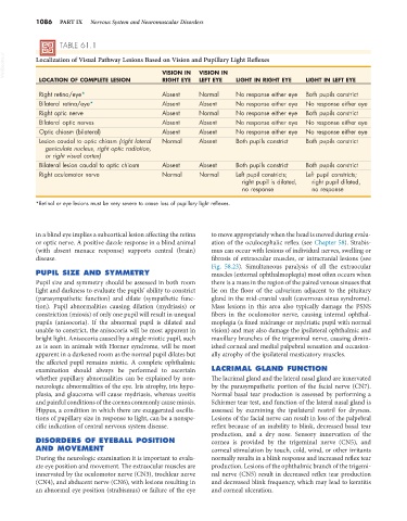
1086   PART IX   Nervous System and Neuromuscular Disorders


                   TABLE 61.1
  VetBooks.ir  Localization of Visual Pathway Lesions Based on Vision and Pupillary Light Reflexes

                                                             VISION IN
                                                  VISION IN
             LOCATION OF COMPLETE LESION          RIGHT EYE  LEFT EYE   LIGHT IN RIGHT EYE   LIGHT IN LEFT EYE
             Right retina/eye*                    Absent     Normal     No response either eye  Both pupils constrict
             Bilateral retina/eye*                Absent     Absent     No response either eye  No response either eye
             Right optic nerve                    Absent     Normal     No response either eye  Both pupils constrict
             Bilateral optic nerves               Absent     Absent     No response either eye  No response either eye
             Optic chiasm (bilateral)             Absent     Absent     No response either eye  No response either eye
             Lesion caudal to optic chiasm (right lateral   Normal  Absent  Both pupils constrict  Both pupils constrict
               geniculate nucleus, right optic radiation,
               or right visual cortex)
             Bilateral lesion caudal to optic chiasm  Absent  Absent    Both pupils constrict  Both pupils constrict
             Right oculomotor nerve               Normal     Normal     Left pupil constricts;   Left pupil constricts;
                                                                          right pupil is dilated,   right pupil dilated,
                                                                          no response          no response

            *Retinal or eye lesions must be very severe to cause loss of pupillary light reflexes.


            in a blind eye implies a subcortical lesion affecting the retina   to move appropriately when the head is moved during evalu-
            or optic nerve. A positive dazzle response in a blind animal   ation of the oculocephalic reflex (see Chapter 58). Strabis-
            (with absent menace response) supports central (brain)   mus can occur with lesions of individual nerves, swelling or
            disease.                                             fibrosis of extraocular muscles, or intracranial lesions (see
                                                                 Fig. 58.23). Simultaneous paralysis of all the extraocular
            PUPIL SIZE AND SYMMETRY                              muscles (external ophthalmoplegia) most often occurs when
            Pupil size and symmetry should be assessed in both room   there is a mass in the region of the paired venous sinuses that
            light and darkness to evaluate the pupils’ ability to constrict   lie on the floor of the calvarium adjacent to the pituitary
            (parasympathetic function) and dilate (sympathetic func-  gland in the mid-cranial vault (cavernous sinus syndrome).
            tion). Pupil abnormalities causing dilation (mydriasis) or   Mass lesions in this  area also typically damage the PSNS
            constriction (miosis) of only one pupil will result in unequal   fibers in the oculomotor nerve, causing internal ophthal-
            pupils  (anisocoria).  If  the abnormal  pupil is  dilated  and   moplegia (a fixed midrange or mydriatic pupil with normal
            unable to constrict, the anisocoria will be most apparent in   vision) and may also damage the ipsilateral ophthalmic and
            bright light. Anisocoria caused by a single miotic pupil, such   maxillary branches of the trigeminal nerve, causing dimin-
            as is seen in animals with Horner syndrome, will be most   ished corneal and medial palpebral sensation and occasion-
            apparent in a darkened room as the normal pupil dilates but   ally atrophy of the ipsilateral masticatory muscles.
            the affected pupil remains miotic. A complete ophthalmic
            examination should always be performed to ascertain   LACRIMAL GLAND FUNCTION
            whether pupillary abnormalities can be explained by non-  The lacrimal gland and the lateral nasal gland are innervated
            neurologic abnormalities of the eye. Iris atrophy, iris hypo-  by the parasympathetic portion of the facial nerve (CN7).
            plasia, and glaucoma will cause mydriasis, whereas uveitis   Normal basal tear production is assessed by performing a
            and painful conditions of the cornea commonly cause miosis.   Schirmer tear test, and function of the lateral nasal gland is
            Hippus, a condition in which there are exaggerated oscilla-  assessed by examining the ipsilateral nostril for dryness.
            tions of pupillary size in response to light, can be a nonspe-  Lesions of the facial nerve can result in loss of the palpebral
            cific indication of central nervous system disease.  reflex because of an inability to blink, decreased basal tear
                                                                 production, and  a  dry nose. Sensory innervation  of  the
            DISORDERS OF EYEBALL POSITION                        cornea is provided by the trigeminal nerve (CN5), and
            AND MOVEMENT                                         corneal stimulation by touch, cold, wind, or other irritants
            During the neurologic examination it is important to evalu-  normally results in a blink response and increased reflex tear
            ate eye position and movement. The extraocular muscles are   production. Lesions of the ophthalmic branch of the trigemi-
            innervated by the oculomotor nerve (CN3), trochlear nerve   nal nerve (CN5) result in decreased reflex tear production
            (CN4), and abducent nerve (CN6), with lesions resulting in   and decreased blink frequency, which may lead to keratitis
            an abnormal eye position (strabismus) or failure of the eye   and corneal ulceration.