
Page 1115 - Small Animal Internal Medicine, 6th Edition
P. 1115
CHAPTER 61 Loss of Vision and Pupillary Abnormalities 1087
LOSS OF VISION gain, and lethargy with clinical, serum biochemical, and uri-
nalysis findings typical of hyperadrenocorticism, but endo-
VetBooks.ir AND OPTIC NERVE crine tests and advanced imaging of the pituitary and adrenal
LESIONS OF THE RETINA, OPTIC DISK,
glands rarely (<20%) confirm that disorder. In the early
Concurrent loss of vision and diminished or absent PLR
bilaterally symmetric retinal degeneration becomes appar-
indicate the presence of a lesion affecting both the visual and stages of SARDS, both fundi appear normal, but with time a
PLR pathways. Unilateral severe lesions of the retina, optic ent, with hyperreflectivity of the tapetal fundus and attenu-
disk, or optic nerve before the optic chiasm result in impaired ation of retinal blood vessels. These retinal changes are
vision and loss of the direct PLR in the affected eye as well indistinguishable from chronic retinal degeneration caused
as a loss of the consensual PLR in the opposite eye when light by other conditions. Early SARDS is differentiated from ret-
is directed into the affected eye (see Table 61.1). The direct robulbar optic neuritis by its extinguished (flat-line) electro-
and consensual response should be normal (both pupils con- retinogram (ERG), demonstrating photoreceptor death.
strict) when light is directed in the unaffected eye. Ocular or Pathogenesis of the disorder is uncertain. Systemic signs are
optic nerve disease must be very severe to cause complete usually transient and resolve without treatment, but the
loss of PLRs. Whenever an animal is evaluated for blindness, blindness is permanent.
the retina should be carefully examined to rule out disorders
such as progressive retinal atrophy, retinal dysplasia, retinal Optic Neuritis
detachment, retinal hemorrhage, and chorioretinitis (Fig. Inflammation of the optic nerves causes blindness and loss
61.4). Optic nerve atrophy secondary to glaucoma or trauma of PLRs. Funduscopic evaluation may reveal optic disk swell-
must also be eliminated as a cause of blindness and PLR loss. ing and discoloration (red) with or without associated retinal
detachment and hemorrhage. When optic neuritis occurs
Sudden Acquired Retinal Degeneration posterior to the globes (i.e., retrobulbar), the visible portion
Sudden acquired retinal degeneration syndrome (SARDS) is of the optic nerves will be normal. In dogs with blindness
a syndrome causing sudden bilateral degeneration of retinal and loss of PLRs with a normal-appearing fundus, ERG is
photoreceptors in dogs. Middle-aged and older dogs of any required to differentiate bilateral retrobulbar optic neuritis
breed can be affected, with females and obese individuals (normal ERG) from SARDS (flat-line ERG).
predisposed. The primary presenting complaint is loss of Optic neuritis is most commonly seen as an isolated
vision, with complete blindness occurring over a period of idiopathic immune-mediated disorder affecting one or both
hours to weeks and often overnight. Pupils are dilated and optic nerves in dogs, but it may also be a manifestation
PLRs are sluggish in dogs examined shortly after vision loss of systemic infectious disease (Box 61.2), especially canine
and absent in dogs with advanced disease. Many affected distemper, tick-borne diseases, fungal infections, and bacte-
dogs have concurrent polyuria, polydipsia, panting, weight rial meningoencephalitis. Noninfectious disorders such as
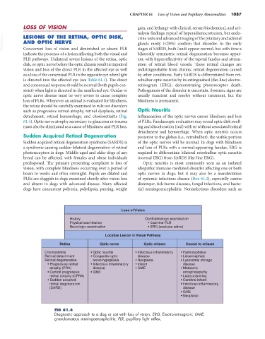
Loss of Vision
History Ophthalmologic examination
Physical examination • Examine PLR
Neurologic examination • ERG (evaluate retina)
Localize Lesion in Visual Pathway
Retina Optic nerve Optic chiasm Caudal to chiasm
Chorioretinitis • Optic neuritis • Infectious inflammatory • Hydrocephalus
Retinal detachment • Congenital optic disease • Lissencephaly
Retinal degeneration nerve hypoplasia • Neoplasia • Lysosomal storage
• Progressive retinal • Infectious inflammatory • Infarct disease
atrophy (PRA) disease • GME • Metabolic
• Central progressive • GME encephalopathy
retinal atrophy (CPRA) • Lead poisoning
• Sudden acquired • Cerebral infarct
retinal degeneration • Infectious inflammatory
(SARD) disease
• GME
• Neoplasia
FIG 61.4
Diagnostic approach to a dog or cat with loss of vision. ERG, Electroretinogram; GME,
granulomatous meningoencephalitis; PLR, pupillary light reflex.