
Page 442 - The Veterinary Laboratory and Field Manual 3rd Edition
P. 442
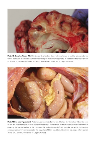
Plate 45 See also Figure A2.2  Bovine cerebral cortex. Note multifocal areas of slightly raised, red areas
                within meninges and extending into the cortical grey matter corresponding to areas of ischaemic infarction
                as a result of bacterial vasculitis. Photo: C. MacGowan, University of Calgary, Canada.


                Plate 46 See also Figure A2.5  Abdomen, cat. Acute inflammation. The last rib (•) and liver (*) can be seen
                on the left side of the picture with loops of intestine (†) in the centre. Note abundant stands of tan fibrin (‡)
                covering the serosal surface of the intestines. Note also the subtle finely granular texture of the intestinal
                serosa which can in some cases be the only sign of fibrin exudation. Abdomen, cat, acute inflammation.
                Photo: Dr J. Davies, University of Calgary, Canada.


       Veterinary-plates.indd   25                                                         26/03/2019   10:14