
Page 523 - Feline diagnostic imaging
P. 523
30.4 Alternate Modalities 535
(a) (b)
(c)
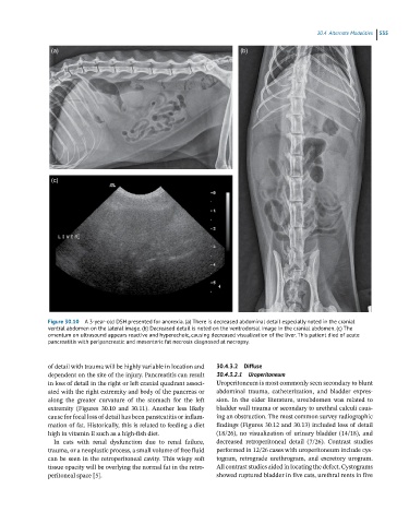
Figure 30.10 A 3-year-old DSH presented for anorexia. (a) There is decreased abdominal detail especially noted in the cranial
ventral abdomen on the lateral image. (b) Decreased detail is noted on the ventrodorsal image in the cranial abdomen. (c) The
omentum on ultrasound appears reactive and hyperechoic, causing decreased visualization of the liver. This patient died of acute
pancreatitis with peripancreatic and mesenteric fat necrosis diagnosed at necropsy.
of detail with trauma will be highly variable in location and 30.4.3.2 Diffuse
dependent on the site of the injury. Pancreatitis can result 30.4.3.2.1 Uroperitoneum
in loss of detail in the right or left cranial quadrant associ- Uroperitoneum is most commonly seen secondary to blunt
ated with the right extremity and body of the pancreas or abdominal trauma, catheterization, and bladder expres-
along the greater curvature of the stomach for the left sion. In the older literature, uroabdomen was related to
extremity (Figures 30.10 and 30.11). Another less likely bladder wall trauma or secondary to urethral calculi caus-
cause for focal loss of detail has been pansteatitis or inflam- ing an obstruction. The most common survey radiographic
mation of fat. Historically, this is related to feeding a diet findings (Figures 30.12 and 30.13) included loss of detail
high in vitamin E such as a high‐fish diet. (18/26), no visualization of urinary bladder (14/18), and
In cats with renal dysfunction due to renal failure, decreased retroperitoneal detail (7/26). Contrast studies
trauma, or a neoplastic process, a small volume of free fluid performed in 12/26 cases with uroperitoneum include cys-
can be seen in the retroperitoneal cavity. This wispy soft togram, retrograde urethrogram, and excretory urogram.
tissue opacity will be overlying the normal fat in the retro- All contrast studies aided in locating the defect. Cystograms
peritoneal space [5]. showed ruptured bladder in five cats, urethral rents in five