
Page 528 - Feline diagnostic imaging
P. 528
(a) (b)
(c)
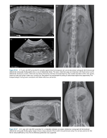
Figure 30.16 A 12-year-old DSH presented for possible gastrointestinal neoplasia. (a) Lateral abdominal radiograph. (b) Ventrodorsal
abdominal radiograph. Radiographs show a loss of abdominal detail. The small intestinal loops are distended with fluid and air. (c) On
abdominal ultrasound, a small intestinal loop shows thickening and loss of normal layering. Fine needle aspiration of the liver, spleen,
intestinal wall, and lymph nodes was inconclusive. The patient was euthanized following an abdominal exploratory laparotomy. The
histopathologic findings were consistent with feline infectious peritonitis.
(b)
(a)
Figure 30.17 A 4.5-year-old male DSH presented for a distended abdomen. (a) Lateral abdominal radiograph. (b) Ventrodorsal
abdominal radiograph. Radiography images shows a large amount of fluid within the peritoneal cavity. The cat was positive for the
feline immunodeficiency virus. Feline infectious peritonitis was suspected.